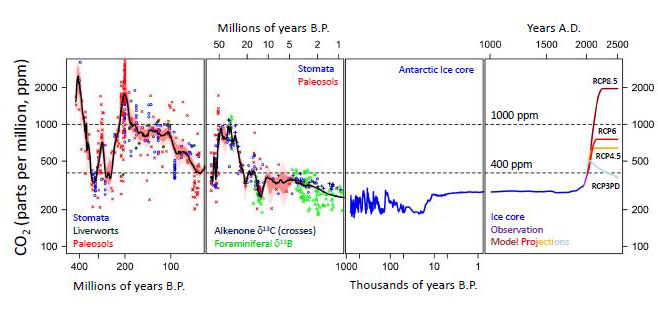
CO2 levels over the past 500 million years. Foster et al – Descent into the icehouse.
Posts made by AlphaCog
-
RE: Study: CO2 may be “bad” for the climate, but is beneficial for humans
-
Improving Metabolic Control Leads to Better Working Memory in Adults With Type 2 Diabetes
OBJECTIVE—The goals of this study were to determine whether improvements in metabolic control can ameliorate the cognitive dysfunction associated with type 2 diabetes and evaluate the possibility that such improvements are mediated by changes in circulating insulin or insulin resistance.
RESEARCH DESIGN AND METHODS—This randomized double-blind trial enrolled 145 subjects at 18 centers in the U.S. Older adults with type 2 diabetes receiving metformin monotherapy received add-on therapy with either rosiglitazone, a thiazolidinedione insulin sensitizer, or glyburide. Cognitive function was assessed at baseline and week 24 using the Digit Symbol Substitution Test, the Rey Auditory Verbal Learning Test, and the Cambridge Neuropsychological Test Automated Battery.
RESULTS—Pretreatment fasting plasma glucose (FPG) in both groups was similar, and after 24 weeks both treatment groups showed similar significant reductions in FPG (2.1–2.3 mmol/l). Working memory improved with both rosiglitazone (P < 0.001) and glyburide (P = 0.017). Improvement (25–31% reduction in errors) was most evident on the Paired Associates Learning Test and was significantly correlated (r = 0.30) with improved glycemic control as measured by FPG.
CONCLUSIONS—Similar and statistically significant cognitive improvement was observed with both rosiglitazone and glyburide therapy, and the magnitude of this effect was correlated with the degree to which FPG improved. These results suggest that a cognitive benefit is achievable with pharmacological interventions targeting glycemic control.
https://diabetesjournals.org/care/article/29/2/345/26200/Improving-Metabolic-Control-Leads-to-Better -
RE: Humans absorb nutrients/chemicals from air, just like they absorb them from food
Would this explain that animals in barren areas such as polar bears and African elephants tended to be larger than their forest counterparts, due to lack of allergens in the air?
-
RE: Gravitational stress and Exercise
Histological and biochemical study of the superficial abdominal fascia and its implication in obesity
Abstract
The advancement of liposculpturing and fascial flaps in reconstructive surgery has renewed interest in the superficial fascia of abdomen. Its histological and biochemical composition may play a vital role in maintaining strength and elasticity of the fascia. Hence, study of abdominal fascia for the elastic, collagen, and hydroxyproline contents is desirable to understand asymmetrical bulges and skin folds and in improving surgical treatment of obesity. Samples of superficial fascia were collected from of upper and lower abdomen from 21 fresh cadavers (15 males and 6 females). Samples were stained using Verhoeff–Van Gieson stain. Digital images of superficial fascia were analyzed using TissueQuant software. The samples were also subjected to hydroxyproline estimation. The superficial fascia was formed by loosely packed collagen fibers mixed with abundant elastic fibers and adipose tissue. Elastic contents and collagen contents of superficial fascia were significantly more in the upper abdomen than that in the lower abdomen in males. Hydroxyproline content of superficial fascia of upper abdomen was significantly more than that of lower abdomen in both males and females. The elastic, collagen and hydroxyproline contents of superficial fascia of upper abdomen were higher compared to the lower abdomen. This may be a reason for asymmetric bulging over abdomen and more sagging fold of skin in the lower abdomen than in the upper abdomen. This study may therefore be helpful in finding new ways to manage obesity and other body contour deformities.
https://pmc.ncbi.nlm.nih.gov/articles/PMC5052227/Muscle Pain: It May Actually Be Your Fascia
Ways to Relieve Fascia Pain
There are various strategies that work to loosen up painful knots, such as:Heat therapy: Apply a heating pad to the affected area or take a warm shower or bath.
Yoga therapy: See a highly trained yoga therapist to get a regimen of yoga poses targeted to treat your area of pain. (Yoga therapy works in the same manner as physical therapy — the therapist creates a routine and you practice it at home between visits.)
Foam rolling: Try a foam roller, a cylinder of hard foam that you roll your body over to release tension. It’s a form of self-massage. You can also do this with a lacrosse ball.
Massage therapy: Schedule multiple therapeutic massage sessions with an experienced therapist who can find and apply pressure to release knots.
Acupuncture: The insertion of acupuncture needles into trigger points can cause tense tissue fibers to relax.
https://www.hopkinsmedicine.org/health/wellness-and-prevention/muscle-pain-it-may-actually-be-your-fasciaYoga Poses for Myofascial Release | 5 Poses to Try
https://www.acefitness.org/resources/everyone/blog/5667/yoga-poses-for-myofascial-release-5-poses-to-try/ -
Gravitational stress and Exercise
Influence of gravity on cardiovascular reflexes from skeletal muscle receptors
Abstract
Skeletal muscles are important reflexogenic areas of the cardiovascular system. The afferent pathways of the reflex loops involve slow-conducting group III and group IV fibers that are excited by mechanical and chemical events in the muscle. The present paper reviews a series of experiments dealing with the question of whether those afferents are also influenced by gravitational forces. The results of these studies suggest the following answers: 1) gravitational forces can modulate cardiovascular reflexes from exercising skeletal muscles. 2) This effect is primarily due to changes in the interstitial fluid volume rather than to a direct mechanical influence, venous pressure, or venous volume. 3) The amplitudes of heart rate and blood pressure responses during exercise are inversely related to the local interstitial volume. Measurements during post-exercise circulatory arrest indicate that this sensitivity is mainly mediated by muscle chemoreceptors. These receptors, which also contribute to the spinal control of movement, generally appear to be sensitized by regional fluid losses and desensitized by overhydration of their environment.
https://pubmed.ncbi.nlm.nih.gov/8897399/Muscle Forces or Gravity: What Predominates Mechanical Loading on Bone?
However, even low-impact gravitational loading may have beneficial skeletal effects, because standing for 40 h·wk−1 or more (which presumably included time spent walking) was associated with a 34% to 46% lower risk of hip fracture (9).
...
several lines of research suggest that physical activities that involve impact forces, and therefore generate both gravitation and muscle loading, are most likely to have beneficial effects on bone metabolism and reduce fracture risk.
https://pmc.ncbi.nlm.nih.gov/articles/PMC3037021/Gravitational Effects on Human Physiology
Abstract
Physical working capacity decreases with age and also in microgravity. Regardless of age, increased physical activity can always improve the physical adaptability of the body, although the mechanisms of this adaptability are unknown. Physical exercise produces various mechanical stimuli in the body, and these stimuli may be essential for cell survival in organisms. The cytoskeleton plays an important role in maintaining cell shape and tension development, and in various molecular and/or cellular organelles involved in cellular trafficking. Both intra and extracellular stimuli send signals through the cytoskeleton to the nucleus and modulate gene expression via an intrinsic property, namely the "dynamic instability" of cytoskeletal proteins. αB-crystallin is an important chaperone for cytoskeletal proteins in muscle cells. Decreases in the levels of αB-crystallin are specifically associated with a marked decrease in muscle mass (atrophy) in a rat hindlimb suspension model that mimics muscle and bone atrophy that occurs in space and increases with passive stretch. Moreover, immunofluorescence data show complete co-localization of αB-crystallin and the tubulin/microtubule system in myoblast cells. This association was further confirmed in biochemical experiments carried out in vitro showing that αB-crystallin acts as a chaperone for heat-denatured tubulin and prevents microtubule disassembly induced by calcium. Physical activity induces the constitutive expression of αB-crystallin, which helps to maintain the homeostasis of cytoskeleton dynamics in response to gravitational forces. This relationship between chaperone expression levels and regulation of cytoskeletal dynamics observed in slow anti-gravitational muscles as well as in mammalian striated muscles, such as those in the heart, diaphragm and tongue, may have been especially essential for human evolution in particular. Elucidation of the intrinsic properties of the tubulin/microtubule and chaperone αB-crystallin protein complex systems is expected to provide valuable information for high-pressure bioscience and gravity health science.
https://pubmed.ncbi.nlm.nih.gov/26174402/Perspective on the Impact of Weightlessness on Calcium and Bone Metabolism
Abstract
As humans venture into space to colonize the moon and travel to distant planets in the 21st century, they will be confronted with a bone disease that could potentially limit their space exploration activities or put them at risk for fracture when they return to earth. It is now recognized that an unloading of the skeleton, either due to strict bed rest or in zero gravity, leads on average to a 1%–2% reduction in bone mineral density at selected skeletal sites each month. The mechanism by which unloading of the skeleton results in rapid mobilization of calcium stores from the skeleton is not fully understood, but it is thought to be related to down regulation in PTH and 1,25-dihydroxyvitamin D3 production. Bone modeling and mineralization in chick embryos is not affected by microgravity, suggesting that bone cells adapt and ultimately become addicted to gravity in order to maintain a structurally sound skeleton. Strategies need to be developed to decrease microgravity-induced bone resorption by either mimicking gravity’s effect on bone metabolism, or enhancing physically or pharmacologically bone formation in order to preserve astronauts’ bone health.
https://www.sciencedirect.com/science/article/pii/S8756328298000143Does gravitational pressure of blood hinder flow to the brain of the giraffe?
The giraffe has a high aortic pressure. This is not for driving the blood to its head but is for minimizing the gravitational drop of intravascular pressure and collapse of the vessels. The cerebral circulation is protected by the cerebrospinal fluid which undergoes parallel changes in pressure with posture. Other vessels in the head are less protected by connective tissue, surrounding muscles and other structures.
https://www.sciencedirect.com/science/article/abs/pii/0300962986905621The roles of exercise in bone remodeling and in prevention and treatment of osteoporosis
Conclusion
Exercise increases bone mineral density, bone mass, bone strength, and bone mechanical properties. It seems to directly or indirectly act on almost all the bone cell types and affect many aspects of bone remodeling.
https://www.sciencedirect.com/science/article/abs/pii/S007961071500228XRecombinant Irisin Prevents the Reduction of Osteoblast Differentiation Induced by Stimulated Microgravity through Increasing β-Catenin Expression
Conclusions: The present study is the first to show that r-irisin positively regulates osteoblast differentiation under simulated microgravity through increasing β-catenin expression, which may reveal a novel mechanism, and it provides a prevention strategy for bone loss and muscle atrophy induced by microgravity.
https://www.mdpi.com/1422-0067/21/4/1259 -
Gastric emptying during walking and running: effects of varied exercise intensity
Summary
Gastric emptying is increased during running (50%–70% maximal aerobic uptake, as compared to rest. Whether this increase varies as a function of mode (i. e. walking vs running) and intensity of treadmill exercise is unknown. To examine the gastric emptying characteristics of water during treadmill exercise performed over a wide range of intensities relative to resting conditions, 10 men ingested 400 ml of water prior to each of six 15 min exercise bouts or 15 min of seated rest. Three bouts of walking exercise (1.57 m · s−1) were performed at increasing grades eliciting ∼28%, 41% or 56% of. On a seperate day, three bouts of running (2.68 ms−1) exercise were performed at grades eliciting ∼57%, 65% or 75% of. Gastric emptying was increased during treadmill exercise at all intensities excluding 75% as compared to rest. Gastric emptying was similar for all intensities during walking and at 57% and 65% during running. However, running at 74% decreased the volume of original drink emptied as compared to all lower exercise intensities. Stomach secretions were markedly less during running as compared to walking and rest. These data demonstrate that gastric emptying is similarly increased during both moderate intensity (∼8%–65%) walking or running exercise as compared to resting conditions. However, gastric emptying decreases during high intensity exercise. Increases in gastric emptying during moderate intensity treadmill exercise may be related to increases in intragastric pressure brought about by contractile activity of the abdominal muscles.
https://link.springer.com/article/10.1007/BF00643522Three 15-min Bouts of Moderate Postmeal Walking Significantly Improves 24-h Glycemic Control in Older People at Risk for Impaired Glucose Tolerance
RESULTS
Both sustained morning walking (127 ± 23 vs. 118 ± 14 mg dL−1) and postmeal walking (129 ± 24 vs. 116 ± 13 mg dL−1) significantly improved 24-h glycemic control relative to the control day (P < 0.05). Moreover, postmeal walking was significantly (P < 0.01) more effective than 45 min of sustained morning or afternoon walking in lowering 3-h postdinner glucose between the control and experimental day.
https://diabetesjournals.org/care/article/36/10/3262/30770/Three-15-min-Bouts-of-Moderate-Postmeal-Walking -
RE: Nighttime melatonin intake increase Vitamin D levels
The role of melatonin in bone regeneration: A review of involved signaling pathways
Abstract
Increasing bone resorption followed by decreasing bone mineralization are hallmarks of bone degeneration, which mostly occurs in the elderly population and post-menopausal women. The use of mesenchymal stem cells (MSCs) has raised many promises in the field of bone regeneration due to their high osteoblastic differentiation capacity and easy availability from abundant sources. A variety of compounds, including growth factors, cytokines, and other internal factors, have been combined with MSCs to increase their osteoblastic differentiation capacity. One of these factors is melatonin, whose possible regulatory role in bone metabolism and formation has recently been suggested by many studies. Melatonin also is a potential signaling molecule and can affect many of the signaling pathways involved in MSCs osteoblastic differentiation, such as activation of PI3K/AKT, BMP/Smad, MAPK, NFkB, Nrf2/HO-1, Wnt, SIRT/SOD, PERK/ATF4. Furthermore, melatonin in combination with other components such as strontium, vitamin D3, and vitamin K2 has a synergistic effect on bone microstructure and improves bone mineral density (BMD). In this review article, we aim to summarize the regulatory mechanisms of melatonin in osteoblastic differentiation of MSCs and underling involved signaling pathways as well as the clinical potential of using melatonin in bone degenerative disorders.
https://www.sciencedirect.com/science/article/abs/pii/S0300908422002127Melatonin-micronutrients Osteopenia Treatment Study (MOTS): a translational study assessing melatonin, strontium (citrate), vitamin D3 and vitamin K2 (MK7) on bone density, bone marker turnover and health related quality of life in postmenopausal osteopenic women following a one-year double-blind RCT and on osteoblast-osteoclast co-cultures
https://pubmed.ncbi.nlm.nih.gov/28130552/ -
Nighttime melatonin intake increase Vitamin D levels
Results: Ten healthy young women between the ages of 20 and 23 were included in our study after obtaining their consent. The mean age of the participants was 21.63 ± 0.94 years. The average height of our patients was 165.72 ± 3.38 m. the average weight of our patients was 59.45 ± 3.5 kg. Vitamin D levels were significantly higher after taking melatonin (mean 13.6 ± 7.9 ng/ml) than after taking placebo (mean 12.68 ± 5.7 ng/ml) during the follicular phase (P=0.001). Whereas, the increase in vitamin D with the intake of melatonin (mean of 15.8 ± 7.8 ng/ml) compared to the taking of placebo (mean of 14.6 ± 5.7 ng/ml) was not significant during the luteal phase (P=0.7). In addition, there was no significant variation in vitamin D levels between the luteal phase and the follicular phase when taking placebo (P= 0.07) and when taking melatonin (P= 0.25).
https://www.endocrine-abstracts.org/ea/0081/ea0081ep634 -
RE: Substantively Lowered Levels of Pantothenic Acid (Vitamin B5) in Several Regions of the Human Brain in Parkinson's Disease Dementia
Pantethine
Because of pantothenic acid’s role in triglyceride synthesis and lipoprotein metabolism, experts have hypothesized that pantothenic acid supplementation might reduce lipid levels in patients with hyperlipidemia [19].Several clinical trials have shown that the form of pantothenic acid known as pantethine reduces lipid levels when taken in large amounts [20], but pantothenic acid itself does not appear to have the same effects [1]. A 2005 review included 28 small clinical trials (average sample size of 22 participants) that examined the effect of pantethine supplements (median daily dose of 900 mg for an average of 12.7 weeks) on serum lipid levels in a total of 646 adults with hyperlipidemia [20]. On average, the supplements were associated with triglyceride declines of 14.2% at 1 month and 32.9% at 4 months. The corresponding declines in total cholesterol were 8.7% and 15.1%, and for low-density lipoprotein (LDL) cholesterol were 10.4% and 20.1%. The corresponding increases in high-density lipoprotein (HDL) cholesterol were 6.1% and 8.4%.
A few additional clinical trials have assessed pantethine’s effects on lipid levels since the publication of the 2005 review. A double-blind trial in China randomly assigned 216 adults with hypertriglyceridemia (204–576 mg/dl) to supplementation with 400 U/day CoA or 600 mg/day pantethine [21]. All participants also received dietary counseling. Triglyceride levels dropped by a significant 16.5% with pantethine compared with baseline after 8 weeks. Concentrations of total cholesterol and non-HDL cholesterol also declined modestly but significantly from baseline. However, these declines might have been due, at least in part, to the dietary counseling that the participants received.
Two randomized, blinded, placebo-controlled studies by the same research group in a total of 152 adults with low to moderate cardiovascular disease risk found that 600 mg/day pantethine for 8 weeks followed by 900 mg/day for 8 weeks plus a therapeutic lifestyle change diet resulted in small but significant reductions in total cholesterol, LDL cholesterol, and non-HDL cholesterol compared with placebo after 16 weeks [19,22]. Increasing the amount of pantethine from 600 to 900 mg/day did not increase the magnitude of reduction in the lipid measures.
Additional studies are needed to determine whether pantethine supplementation has a beneficial effect on hyperlipidemia independently of, and together with, eating a heart-healthy diet. Research is also needed to determine the mechanisms of pantethine’s effects on lipid levels.
https://ods.od.nih.gov/factsheets/PantothenicAcid-HealthProfessional/#h9 -
RE: Tea consumption increases bone density and reduces the risk of osteoporosis
@yerrag It's not the calcium amount but the stress-reducing bioactive compounds in tea having a positive effect on metabolism of bone/glucose.
-
RE: Tea consumption increases bone density and reduces the risk of osteoporosis
Monitoring fluorine levels in tea leaves from major producing areas in China and the relative health risk
Abstract
The trace element fluoride can exert a preventive function on dental caries at moderate intake levels, but can also lead to skeletal fluorosis and cognitive impairment when consumed in excessive amounts. Tea is a fluoride hyperaccumulator, yet the risks of fluoride intake via tea consumption are yet to be investigated. In this study, we measured fluoride levels in common tea products from major cultivation provinces in China. Fluoride contents ranged from 6 to 808 mg/kg. Of the samples, 15.6% (34/218) demonstrated fluoride contents which exceeded the maximum residue limit of 200 mg/kg. The samples with high fluoride contents mainly represented oolong tea and dark tea. Fluoride contents in tea were significantly varied between producing regions, which is mainly due to different degree of soil acidification and fluoride occurrence in groundwater. Daily fluoride intake via tea consumption among the general population ranged from 0.02 to 2.20 mg/day, which is lower than the tolerable upper fluoride intake level (UL) of 3.50 mg/day based on Chinese Dietary Reference Intakes, but rather close to the UL of 2.50 mg/day set by the European Food Safety Authority. A risk assessment indicated that the consumption of tea was not associated with any significant health risks (hazard quotient ≤ 0.54). However, we found a potential risk for the simultaneous consumption of tea, drinking water, wheat and seafood based on a simulation assessment. Fluoride intake among the high-risk population should be continuously monitored.
https://www.sciencedirect.com/science/article/abs/pii/S0889157523000790The effects of green tea consumption on metabolic and anthropometric indices in patients with Type 2 diabetes
Conclusion:
Drinking four cups of green tea led to a significant reduction in weight and systolic blood pressure.
https://pmc.ncbi.nlm.nih.gov/articles/PMC3908530/Shaded Japanese Green Teas
Shaded and unshaded teas refer to how the tea is cultivated in Japan. Shading, or kabuse, is done to reduce the sunlight that the tea plant receives by about 85%, which lowers the plant’s photosynthesis.To put it succinctly, shading is done to keep the L-theanine content high, and the formation of catechins low. L-theanine is responsible for the umami taste, whereas catechins are responsible for the tea tasting astringent or bitter.
Additionally, shading produces a sweeter leaf because of the higher sugar content. It also tends to create darker and greener leaves due to the plant creating more chlorophyll.
Shading is generally done for a short period (2 to 4 weeks) before the tea leaves are picked. For example, Kabuse Sencha is shaded for 2 weeks, Gyokuro for 3, and finally, Matcha for 4 weeks. Shading is done either directly on the top of the plant or more traditionally with shelf style shading. Shading is also only done in Japan.
https://obubutea.com/comparing-shaded-japanese-green-teas/ -
Tea consumption increases bone density and reduces the risk of osteoporosis
Results:
Our study was based on 17 journal articles, including 2 prospective cohort studies, 4 case–control studies, and 11 cross-sectional studies. In the present study, the total OR of osteoporosis for the highest versus the lowest categories of tea consumption was 0.62 (95% CI, 0.46–0.83), with significant heterogeneity among studies (I2 = 94%, P < .01). There was, however, no publication bias of the meta-analysis about tea consumption and osteoporosis. Subgroup analysis showed that tea consumption could reduce the risk of osteoporosis in all examined subgroups.Conclusion:
In the present study, it can be concluded from the results that tea consumption can reduce the risk of osteoporosis. -
RE: The benefits of smoking, if any, may be due to lowering endotoxin and estrogen
Ongoing trial:
Efficacy of Nicotine in Preventing COVID-19 Infection (NICOVID-PREV)
https://clinicaltrials.gov/study/NCT04583410 -
RE: Fasting-induced autophagy versus saturated-fat-induced apoptosis.
My poll was voted on and I forgot to check:
https://lowtoxinforum.com/threads/oxidized-cholesterol-vs-pufa.28333/Which one is worse? Oxidized Cholesterol vs Polyunsaturated Fatty Acid
- Oxidized Cholesterol Votes: 2
- Polyunsaturated Fatty Acid Votes: 7
-
Fasting-induced autophagy versus saturated-fat-induced apoptosis.
Answered my question from 5 years ago with ChatGPT.
https://lowtoxinforum.com/threads/unsaturated-fat-protects-against-saturated-fat-induced-lipotoxicity.28418/post-427668GPT-40:
Fasting-induced autophagy and saturated-fat-induced apoptosis are two distinct processes that play crucial roles in cellular health, but they have very different effects and mechanisms.1. Fasting-Induced Autophagy
- Mechanism: Autophagy is a natural, regulated process of breaking down and recycling damaged or dysfunctional cellular components. When fasting, nutrient availability (particularly glucose and amino acids) drops, activating signaling pathways (e.g., AMPK and mTOR inhibition) that trigger autophagy. Cells then begin to digest damaged proteins, organelles, and other cellular debris, providing energy and essential building blocks for cellular repair and rejuvenation.
- Benefits: Autophagy is associated with various health benefits, including improved metabolic health, protection against neurodegenerative diseases, and potentially increased longevity. By recycling cellular components, autophagy helps cells stay functional and resilient under stress, such as during nutrient scarcity.
- Protective Effects: Autophagy supports cellular survival by removing potentially toxic cellular materials and promoting energy efficiency, acting as a self-preservation mechanism.
2. Saturated-Fat-Induced Apoptosis
- Mechanism: Apoptosis is a programmed cell death pathway that helps eliminate damaged or unneeded cells. Saturated fats, especially when consumed in high amounts, can trigger apoptosis by causing oxidative stress, inflammation, and mitochondrial dysfunction. Excess saturated fats can disrupt the lipid composition of cell membranes and impair cellular function, leading to stress signals that activate apoptosis through pathways like caspase activation and mitochondrial damage.
- Drawbacks: While apoptosis is a healthy and necessary process for removing damaged cells, chronic exposure to saturated fats can increase the rate of cell death in vital tissues like the liver, pancreas, and even certain areas of the brain. This has been linked to insulin resistance, fatty liver disease, and increased cardiovascular risks, particularly in the context of a high-saturated-fat diet with low intake of protective nutrients.
- Potential for Damage: In cases of excessive saturated fat consumption, apoptosis can contribute to tissue damage and inflammation, as dying cells release pro-inflammatory signals, potentially harming healthy surrounding cells.
Summary: Fasting-Induced Autophagy vs. Saturated-Fat-Induced Apoptosis
- Autophagy (from fasting) is a beneficial cellular cleaning and repair process, supporting cellular function and resilience, particularly under nutrient-scarce conditions.
- Apoptosis (from excess saturated fat), while protective in certain contexts, can lead to inflammation and tissue damage when triggered excessively. This can compromise tissue health and overall metabolic stability.
In summary, fasting-induced autophagy supports cellular health and longevity by clearing damaged components, while saturated-fat-induced apoptosis is a response to cellular stress that, when chronic, can contribute to metabolic dysfunction and tissue damage.
-
RE: Substantively Lowered Levels of Pantothenic Acid (Vitamin B5) in Several Regions of the Human Brain in Parkinson's Disease Dementia
Its name comes from the Greek πάντοθεν pantothen, meaning "from everywhere", because pantothenic acid, at least in small amounts, is in almost all foods. Deficiency of pantothenic acid is very rare in humans.
https://en.wikipedia.org/wiki/Pantothenic_acidFrom this we infer that people with dementia probably had poor digestion.
Gastrointestinal Diseases, Genetic Risk, and Incident Dementia: A Prospective Cohort Study in 352,463 Middle-Aged Adults
Conclusions and relevance This study found associations of several gastrointestinal diseases with an increased risk of incident dementia, especially early-onset dementia. Our findings imply the great importance of dementia prevention among patients with gastrointestinal diseases.
https://www.medrxiv.org/content/10.1101/2022.11.28.22282820v1.fullDementia with Lewy bodies (DLB) patients suffer more frequently from gastrointestinal dysfunction
https://wfneurology.org/activities/news-events/archived-news/2017-09-18-wcn-dlb-patients-suffer-more-from-gastrointestinal-dysfunctionDysbiosis, gut barrier dysfunction and inflammation in dementia: a pilot study
https://bmcgeriatr.biomedcentral.com/articles/10.1186/s12877-020-01644-2 -
Substantively Lowered Levels of Pantothenic Acid (Vitamin B5) in Several Regions of the Human Brain in Parkinson's Disease Dementia
Abstract
Pantothenic acid (vitamin B5) is an essential trace nutrient required for the synthesis of coenzyme A (CoA). It has previously been shown that pantothenic acid is significantly decreased in multiple brain regions in both Alzheimer's disease (ADD) and Huntington's disease (HD). The current investigation aimed to determine whether similar changes are also present in cases of Parkinson's disease dementia (PDD), another age-related neurodegenerative condition, and whether such perturbations might occur in similar regions in these apparently different diseases. Brain tissue was obtained from nine confirmed cases of PDD and nine controls with a post-mortem delay of 26 h or less. Tissues were acquired from nine regions that show high, moderate, or low levels of neurodegeneration in PDD: the cerebellum, motor cortex, primary visual cortex, hippocampus, substantia nigra, middle temporal gyrus, medulla oblongata, cingulate gyrus, and pons. A targeted ultra-high performance liquid chromatography-tandem mass spectrometry (UHPLC-MS/MS) approach was used to quantify pantothenic acid in these tissues. Pantothenic acid was significantly decreased in the cerebellum (p = 0.008), substantia nigra (p = 0.02), and medulla (p = 0.008) of PDD cases. These findings mirror the significant decreases in the cerebellum of both ADD and HD cases, as well as the substantia nigra, putamen, middle frontal gyrus, and entorhinal cortex of HD cases, and motor cortex, primary visual cortex, hippocampus, middle temporal gyrus, cingulate gyrus, and entorhinal cortex of ADD cases. Taken together, these observations indicate a common but regionally selective disruption of pantothenic acid levels across PDD, ADD, and HD.
https://pubmed.ncbi.nlm.nih.gov/34564384/Localized Pantothenic Acid (Vitamin B5) Reductions Present Throughout the Dementia with Lewy Bodies Brain
Abstract
Background: Localized pantothenic acid deficiencies have been observed in several neurodegenerative diseases such as Alzheimer's disease (AD), Parkinson's disease dementia (PDD), and Huntington's disease (HD), indicating downstream energetic pathway perturbations. However, no studies have yet been performed to see whether such deficiencies occur across the dementia with Lewy bodies (DLB) brain, or what the pattern of such dysregulation may be.Objective: Firstly, this study aimed to quantify pantothenic acid levels across ten regions of the brain in order to determine the localization of any pantothenic acid dysregulation in DLB. Secondly, the localization of pantothenic acid alterations was compared to that previously in AD, PDD, and HD brains.
Methods: Pantothenic acid levels were determined in 20 individuals with DLB and 19 controls by ultra-high performance liquid chromatography-tandem mass spectrometry (UHPLC-MS/MS) across ten brain regions. Case-control differences were determined by nonparametric Mann-Whitney U test, with the calculation of S-values, risk ratios, E-values, and effect sizes. The results were compared with those previously obtained in DLB, AD, and HD.
Results: Pantothenic acid levels were significantly decreased in six of the ten investigated brain regions: the pons, substantia nigra, motor cortex, middle temporal gyrus, primary visual cortex, and hippocampus. This level of pantothenic acid dysregulation is most similar to that of the AD brain, in which pantothenic acid is also decreased in the motor cortex, middle temporal gyrus, primary visual cortex, and hippocampus. DLB appears to differ from other neurodegenerative diseases in being the only of the four to not show pantothenic acid dysregulation in the cerebellum.
Conclusions: Pantothenic acid deficiency appears to be a shared mechanism of several neurodegenerative diseases, although differences in the localization of this dysregulation may contribute to the differing clinical pathways observed in these conditions.
Plain language summary
Decreases in a molecule called pantothenic acid (also known as vitamin B5) have been observed in several areas of the brain in multiple dementia disease, including Alzheimer’s disease, Parkinson’s disease dementia, and Huntington’s disease. However, it is unknown whether such changes also occur in another dementia disease, dementia with Lewy bodies, which shows many of the same symptoms and molecular changes as these conditions. As such, this study was performed in order to determine if and where changes in pantothenic acid occur throughout the dementia with Lewy bodies brain. Using a methodology called liquid chromatography–mass spectrometry, which is able to measure pantothenic acid levels in a highly precise manner in brain tissues, we found that several regions of the dementia with Lewy bodies brain show decreases in pantothenic acid, including some involved in movement such as the substantia nigra and motor cortex, as well as regions associated with cognition and memory such as the hippocampus—looking most similar to the pattern of changes already seen in Alzheimer’s disease. It is possible that these changes contribute to the progression of dementia with Lewy bodies; however, further studies need to be performed to determine at what point these changes happen during the disease and how they may contribute to the development of symptoms. -
RE: Metformin reduces gluconeogenesis that is typical of "fight or flight" stress.
Could Alzheimer’s disease be a maladaptation of an evolutionary survival pathway mediated by intracerebral fructose and uric acid metabolism?
Although biological effects of fructose metabolism and its byproduct, intracellular uric acid, appear critical for the survival of many animals in nature, including our ancestors, in modern society, it appears to be overengaged, increasing the risk for metabolic syndrome, obesity, diabetes, and other conditions
...
uric acid translocatess NADPH oxidase (nicotinamide adenine dinucleotide phosphate oxidase) to the mitochondria, leading to oxidative stress that blocks the citric acid cycle (via inhibition of aconitase) and fatty acid β-oxidation. As mitochondrial function slows, glycolysis takes over, while uric acid inhibits AMP-activated protein kinase, reducing the ability to recover ATP. The effect is a reduction in ATP in the cell, activating a survival switch that includes hunger, thirst, foraging, fat accumulation, and insulin resistance.
...
Uric acid translocates NADPH oxidase (nicotinamide adenine dinucleotide phosphate oxidase) to the mitochondria, where it causes oxidative stress, reducing fatty acid oxidation (blocking enoyl CoA hydratase) while inhibiting aconitase in the citric acid cycle
...
In nature, dietary fructose from excessive intake of fruit provides a major pathway to activate this survival response, much like what occurs in the autumn when bears prepare for hibernation. However, fructose is also produced in the body via the polyol pathway, in which glucose is converted to fructose. The rate-limiting enzyme in the polyol pathway is aldose reductase, and its activity is stimulated during times of stress, such as when nutrient delivery is impaired (hypoxia or ischemia), when water supplies are low (dehydration, hyperglycemia, and hyperosmolarity), or when uric acid levels are high (reflecting degradation of nucleotides and ATP, suggestive of an energy crisis)
...
However, the studies that evaluated the differences between fructose and glucose in cerebral metabolism using BOLD MRI were performed early (∼15 min), thus making it more likely to reflect true differences between fructose and glucose. The striking finding from these studies was that fructose reduced blood flow to the posterior cingulate cortex, the hippocampus, the thalamus, and the occipital cortex; however, blood flow increased to the area of the visual cortex associated with food reward. Cortical blood flow also decreased. Fructose administration also stimulated hunger and desire for food. These responses are consistent with a stimulation of the foraging response. In contrast, glucose inhibited blood flow to the hypothalamus, thalamus, insula, anterior cingulate, and striatum while stimulating blood flow to the cortex. These responses are expected to inhibit not only the foraging response but also responses involving appetite and reward.
...
The brain can generate and metabolize fructose
Our hypothesis suggests that local fructose generation and metabolism may be the critical factor for how fructose induces AD because under normal circumstances, only 1%–2% of ingested fructose reaches the brain. Indeed, the brain is capable of producing fructose. As mentioned earlier, simply raising blood glucose levels increases brain fructose levels in healthy humans. Raising serum osmolality in mice by dehydration or salty food also stimulates fructose production in the brain (hypothalamus). Dietary fructose may also increase fructose production in the brain, possibly by raising uric acid levels in the brain. For example, acutely raising serum uric acid increases uric acid in both the hypothalamus and the hippocampus in association with local inflammation. In turn, uric acid stimulates fructose production and metabolism.
...
Fructose is elevated in the brain of patients with AD
Sorbitol and fructose levels (both components of the polyol pathway) were significantly elevated, averaging 3–5-fold higher in all regions of the brain studied, including the hippocampus, entorhinal cortex, middle temporal gyrus, cingulate cortex, sensory and motor cortex, and cerebellum.
...
Numerous studies have reported that subjects with AD have low serum uric acid levels, suggesting that this might be important to the pathogenesis. However, although serum uric acid may reflect fructose metabolism, it also is a general marker of nutrition status. Clinical manifestations of AD are often preceded by significant weight loss. Which may account for the lower serum uric acid levels on presentation of AD. This may also explain why obesity predicts AD in midlife but actually protects from AD late in life
...
https://www.sciencedirect.com/science/article/pii/S0002916523000047 -
RE: Metformin reduces gluconeogenesis that is typical of "fight or flight" stress.
@LucH Thanks for the link. I'm curious regarding the interpretation of Beta-Amyloid and Tau Proteins by various parties. The behaviour of the proteins seemed to have commonality with Uric Acid.
Metformin prevents p-tau and amyloid plaque deposition and memory impairment in diabetic mice
Abstract
Insulin deficiency or resistance can promote dementia and hallmarks of Alzheimer's disease (AD). The formation of neurofibrillary tangles of p-TAU protein, extracellular Aβ plaques, and neuronal loss is related to the switching off insulin signaling in cognition brain areas. Metformin is a biguanide antihyperglycemic drug used worldwide for the treatment of type 2 diabetes. Some studies have demonstrated that metformin exerts neuroprotective, anti-inflammatory, anti-oxidant, and nootropic effects. This study aimed to evaluate metformin's effects on long-term memory and p-Tau and amyloid β modulation, which are hallmarks of AD in diabetic mice. Swiss Webster mice were distributed in the following experimental groups: control; treated with streptozotocin (STZ) that is an agent toxic to the insulin-producing beta cells; STZ + metformin 200 mg/kg (M200). STZ mice showed significant augmentation of time spent to reach the target box in the Barnes maze, while M200 mice showed a significant time reduction. Moreover, the M200 group showed reduced GFAP immunoreactivity in hippocampal dentate gyrus and CA1 compared with the STZ group. STZ mice showed high p-Tau levels, reduced p-CREB, and accumulation of β-amyloid (Aβ) plaque in hippocampal areas and corpus callosum. In contrast, all these changes were reversed in the M200 group. Protein expressions of p-Tau, p-ERK, pGSK3, iNOS, nNOS, PARP, Cytochrome c, caspase 3, and GluN2A were increased in the parietal cortex of STZ mice and significantly counteracted in M200 mice. Moreover, M200 mice also showed significantly high levels of eNOS, AMPK, and p-AKT expression. In conclusion, metformin improved spatial memory in diabetic mice, which can be associated with reducing p-Tau and β-amyloid (Aβ) plaque load and inhibition of neuronal death.
https://pubmed.ncbi.nlm.nih.gov/34283253/Metformin attenuates Alzheimer's disease-like neuropathology in obese, leptin-resistant mice
Abstract
Diabetes increases the risk of Alzheimer's disease (AD). The pathological hallmarks for AD brains are extracellular amyloid plaques formed by β-amyloid peptide (Aβ) and intracellular neurofibrillary tangles consisting of hyperphosphorylated tau protein. This study was designed to determine AD-like brain changes in mice modeling for type 2 diabetes. The effects of metformin on these changes also were studied. Seven-week old male db/db mice received intraperitoneal injection of 200 mg kg− 1 d− 1 metformin for 18 weeks. They were subjected to Barnes maze at an age of 21 weeks and fear conditioning at an age of 24 weeks to assess their cognitive functions. Hippocampus was harvested after these tests for biochemical evaluation. The db/db mice had more tau phosphorylated at S396 and total tau in their hippocampi than their non-diabetic control db + mice. Activated/phosphorylated c-jun N-terminal kinase (JNK), a tau kinase, was increased in the db/db mouse hippocampus. Metformin attenuated the increase of total tau, phospho-tau and activated JNK. The db/db mice had increased Aβ levels. Metformin attenuated the reduction of synaptophysin, a synaptic protein, in the db/db mouse hippocampus. Metformin did not attenuate the impairments of spatial learning and memory as well as long-term hyperglycemia in the db/db mice. Our results suggest that the db/db mice have multiple AD-like brain changes including impaired cognitive functions, increased phospho-tau and Aβ as well as decreased synaptic proteins. Activation of JNK may contribute to the increased phospho-tau in the db/db mice. Metformin attenuates AD-like biochemical changes in the brain of these mice.
https://www.sciencedirect.com/science/article/abs/pii/S0091305712000640Gout risk in adults with pre-diabetes initiating metformin
Conclusions: Metformin use was associated with a reduced risk of gout among adults with pre-diabetes, suggesting that metformin may be important in lowering gout risk in individuals with pre-diabetes.
https://ard.bmj.com/content/early/2024/05/15/ard-2024-225652Metformin Lowers Serum Urate Levels but Not Gout Risk
https://www.rheumatologyadvisor.com/news/metformin-lowers-serum-urate-but-not-gout-risk/Effect of metformin use on clinical outcomes and serum urate in gout patients with diabetes mellitus: a retrospective cohort study
In conclusion we could not confirm a clinically relevant anti-inflammatory or urate lowering effect of metformin in patients starting ULT treatment and receiving usual care flare prophylaxis.
https://www.ncbi.nlm.nih.gov/pmc/articles/PMC9153141/Metformin Initiation Cuts Gout Risk in Prediabetes
Metformin use lowers the risk for gout by 32% in individuals with prediabetes; however, the treatment doesn't change serum urate or C-reactive protein levels.
https://www.medscape.com/viewarticle/metformin-initiation-cuts-gout-risk-prediabetes-2024a1000a4jMetformin ameliorates high uric acid-induced insulin resistance in skeletal muscle cells
https://www.sciencedirect.com/science/article/abs/pii/S0303720716305482Metformin protects against insulin resistance induced by high uric acid in cardiomyocytes via AMPK signaling pathways in vitro and in vivo
https://www.biorxiv.org/content/10.1101/2021.01.29.428905v1 -
RE: Metformin reduces gluconeogenesis that is typical of "fight or flight" stress.
Fasting enhances extinction retention and prevents the return of fear in humans
Fear is prone to return following extinction that is the basis of exposure therapy for fear-related disorders. Manipulations that enhance the extinction process can be beneficial for treatment. Animal studies have shown that fasting or caloric restriction can enhance extinction and inhibit the return of fear. The present study examined the effects of fasting on fear acquisition, extinction, and the return of fear in humans. One hundred and twenty-five male participants were randomized into a fasting group and food group and exposed to a Pavlovian fear conditioning paradigm. Changes in plasma cortisol and ghrelin levels were examined using enzyme-linked immunosorbent assays. One-night fasting had no effect on fear acquisition but enhanced fear extinction retention and prevented the return of fear, and this effect persisted for at least 6 months. This procedure was also effective for remote fear memory. Plasma ghrelin levels were elevated after fasting and had a negative relationship with the fear response in spontaneous recovery test. However, overnight fasting did not affect cortisol levels. These findings indicate that fasting enhances extinction retention and prevents the return of fear, without influencing fear memory formation. We propose that this novel procedure may open new avenues for promoting extinction-based therapies for fear-related disorders.
https://www.ncbi.nlm.nih.gov/pmc/articles/PMC6177454/The Association of Posttraumatic Stress Disorder with Fast Food and Soda Consumption and Unhealthy Weight Loss Behaviors Among Young Women
Results
PTSD symptoms were associated with an increased frequency of consumption of fast food and soda as well as unhealthy dieting behaviors but not with increased body mass index (BMI).
https://www.ncbi.nlm.nih.gov/pmc/articles/PMC3153863/