Essential Fatty Acids (Omega-3 & Omega-6) are direct building blocks for the skin's lipid bilayers, reducing transepidermal water loss (TEWL).
-
Transepidermal water loss (TEWL), dehydration, and metabolic acidosis are clinically connected, most commonly in the context of severe burn injuries or major skin barrier damage. A breakdown of the skin causes water to passively evaporate, which reduces blood volume, lowers tissue perfusion, and leads to acidic blood.
Changes in Transepidermal Water Loss and Skin Hydration according to Expression of Aquaporin-3 in Psoriasis
https://pubmed.ncbi.nlm.nih.gov/22577267/The repair of impaired epidermal barrier function in rats by the cutaneous application of linoleic acid
https://onlinelibrary.wiley.com/doi/abs/10.1111/j.1365-2133.1976.tb04336.xAnti-Inflammatory and Skin Barrier Repair Effects of Topical Application of Some Plant Oils
https://doi.org/10.3390/ijms19010070Changes in transepidermal water loss and the composition of epidermal lecithin after applications of pure fatty acid triglycerides to the skin of essential fatty acid-deficient rats.
https://doi.org/10.1111/j.1365-2133.1976.tb07012.xKrill oil supplementation improves transepidermal water loss, hydration and elasticity of the skin in healthy adults: Results from two randomized, double-blind, placebo-controlled, dose-finding pilot studies
https://pubmed.ncbi.nlm.nih.gov/39169540/Diet and skin barrier: The role of dietary interventions on skin barrier function.
https://doi.org/10.5826/dpc.1101a132The role of linoleic acid in skin and hair health: A review.
https://www.mdpi.com/1422-0067/26/1/246Feeding the Skin Barrier: The Impact of Macro‐ and Micronutrients on Skin Barrier Function
https://pmc.ncbi.nlm.nih.gov/articles/PMC12626168/Skin pH–dependent Staphylococcus aureus abundance as predictor for increasing atopic dermatitis severity
https://onlinelibrary.wiley.com/doi/full/10.1111/all.14461 -
Mead acid (Omega-9) cannot build fully functional skin lipid bilayers and acts as a biochemical SOS signal. When your body experiences an Essential Fatty Acid Deficiency (EFAD), it tries to compensate by substituting missing omega-6 linoleic acid with de novo synthesized omega-9 Mead acid. [1, 2, 3]
However, this biological substitution fails to restore proper barrier physics, causing the stratum corneum to become leaky, brittle, and hyperproliferative. [3]The Molecular Mismatch: Why Mead Acid Fails
The structural failure of a skin lipid bilayer built with Mead acid comes down to a precise molecular mismatch:
- The Missing Geometry: Linoleic acid (omega-6) has a specific shape and chain length optimized to interlock tightly within acylceramides. This tight packing creates a highly ordered, impermeable orthorhombic crystalline phase.
- The Fluid Kink: Mead acid (omega-9) features three double bonds that introduce a different structural "kink" into the fatty acid tail. It is physically incapable of packing tightly with neighboring ceramides and cholesterol. [2, 4, 5]
- Disrupted Periodicity: Instead of forming neat, tightly packed lamellar sheets (bilayers), Mead acid forces the lipid matrix into a disorganized, more fluid liquid-crystalline phase. [6]
The Consequences of a "Mead Acid Barrier"
When Mead acid replaces linoleic acid in the skin's lipid matrix, the downstream clinical outcomes are highly predictable: [1, 7]
[Linoleic Acid Depletion]
│
▼
[Compensatory Mead Acid Synthesis]
│
▼
[Disorganized, Fluid Lipid Bilayers]
│
├─► High Transepidermal Water Loss (TEWL) ──► Chronic Dryness & Scaling
└─► Compromised Structural Integrity ──► Epidermal Hyperproliferation- Spike in Transepidermal Water Loss (TEWL): Because the Mead acid-substituted lipid bilayers are structurally loose, water easily slips through the intercellular spaces. The skin suffers from severe, unquenchable dehydration regardless of topical humectant use. [3, 7, 8]
- Epidermal Hyperproliferation: Healthy bilayers signal the skin that its shield is intact. The disorganized Mead acid barrier constantly signals a breach, forcing the epidermis to rapidly produce more keratinocytes. This creates the hallmark thick, scaly, and peeling skin seen in EFAD. [3, 8, 9, 10]
- The Triene-to-Tetraene Ratio: In laboratory diagnostics, a high ratio of trienes (Mead acid) to tetraenes (arachidonic acid)—specifically a Holman Index > 0.2—is the definitive biochemical marker used to diagnose systemic essential fatty acid deficiency. [8, 11]
The Therapeutic Divergence
While Mead acid is a poor structural surrogate for your skin's mechanical barrier, emerging research shows it acts as an endogenous anti-inflammatory agent. Topically, Mead acid has been shown to downregulate p38 MAPK pathways and activate PPAR-α, which can actually suppress irritant contact dermatitis or retinoid-induced skin irritation. [2, 12, 13]
However, to truly repair the mechanical, waterproof barrier of the skin, Mead acid must be replaced by reintroducing true omega-6 linoleic acid via systemic nutrition or targeted topical formulations. [14]
Would you like to review:- The exact biochemical pathway where oleic acid converts into Mead acid?
- The optimal topical ratio of linoleic acid to cholesterol for barrier correction?
- How to calculate the triene-to-tetraene ratio from a lipid panel?
- [1] https://www.sciencedirect.com
- [2] https://pmc.ncbi.nlm.nih.gov
- [3] https://lpi.oregonstate.edu
- [4] https://www.instagram.com
- [5] https://pmc.ncbi.nlm.nih.gov
- [6] https://www.sciencedirect.com
- [7] https://www.nbflanes.com
- [8] https://aspenjournals.onlinelibrary.wiley.com
- [9] https://www.shankara.in
- [10] https://pmc.ncbi.nlm.nih.gov
- [11] https://www.jaad.org
- [12] https://pubmed.ncbi.nlm.nih.gov
- [13] https://pmc.ncbi.nlm.nih.gov
- [14] https://curology.com
-
@AlphaZance hi, have you read the burr experiment?
-
It’s impossible to eat a decent diet without getting some PUFA.
There is a tremendous prejudice in the literature favoring the “essential” fatty acids.
Omega 6 encourages sunburn and sun damage in my observation. The lower the PUFA, the better my skin seems to be.
I think Dr. Peat was correct, but I appreciate other views too, so thank you.
-
It’s impossible to eat a decent diet without getting some PUFA.
There is a tremendous prejudice in the literature favoring the “essential” fatty acids.
Omega 6 encourages sunburn and sun damage in my observation. The lower the PUFA, the better my skin seems to be.
I think Dr. Peat was correct, but I appreciate other views too, so thank you.
HI, have you read on Rpf the differents reports of skin worsening on low pufa diet? Dry skin sometimes extremly dry skin is mentionned, and some people improve it by consuming straight seeds oil. Low pufa diet don't necessary lead to skin problems, yet in some cases it can be associated to it and seed oil and/or higher mufa/pufa intake can improve the condition
-
@user1 yes I have read them. I’m not sure what the reason is, or what combination leads to extremely dry skin, but I suspect PUFA makes things worse in the longer run.
-
@user1 yes I have read them. I’m not sure what the reason is, or what combination leads to extremely dry skin, but I suspect PUFA makes things worse in the longer run.
If you stay in the sun with higher propency to burning and skin damage, yes on the long run it might be worst, if you avoid sun exposure it might not. Been wondering if it's possible people with higher mufa/pufa intake either have higher skin moisture and/or sebum at baseline, and at lower temperature set point, and people that are truly more saturated have lower skin moisture and/or sebum at baseline or might need higher temperature set point or sun exposure to be more moisturized
-
Krill oil supplementation improves transepidermal water loss, hydration and elasticity of the skin in healthy adults
Useful omega 6 for skin repair or omega 3 to dampen an immune response
Excerpt from my post: Immunosuppressive effects of omega-3
How can unrefined / unadulterated omega-3 be successfully supplied without increasing oxidative risks through peroxidation? That is the crux of the problem. (1)
Useful omega-6 for skin repair
Yes, hydration and elasticity of the skin in healthy adults is observed when taking evening primrose oil for men or borage oil for women, incredibly rich in gamma-linolenic acid (GLA), which the body metabolizes into dihomo-gamma-linolenic acid (DGLA) to produce powerful anti-inflammatory prostaglandins. This makes it highly effective for soothing dry, irritated skin, reducing fine lines, and restoring the skin's moisture barrier. Dixit AI.
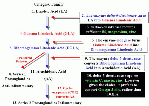
Not sure the best pathway will be reached, but it could be attempted. Too long to explain here. See the figure: LA => GLA => DGLA => AA or PG-2
Studies on the beneficial effects of omega-3s
No one disputes the benefits of an omega-3 regimen against inflammation or in case of an excessive immune response, particularly with EPA, very functional.
Okay, omega-3s are immuno-modulators. However, this applies either in the short term (cure) or when the diet is balanced (specifically, with an omega-3 to omega-6 ratio ranging from 1:1 to 1:4, or at a maximum of 1:10 at some time if limited in time). The US ratio is usually at 1/20 (with food manufactured, or with livestock fed partly on corn meal and soy, instead of grazing for nine months out of twelve).
Anything exceeding the body's requirements will be stored.
At that point, the eicosanoids exert a hormonal effect. It becomes a ticking time bomb (driven by what is known as the arachidonic acid cascade).
What is meant by intake exceeding physiological needs?
The requirements are very weak. And not specifically every day. Omega-3s (ALA) are conditionally essential. This means the body can do without them if the necessary cofactors are present (2). As for omega-6s, AA and DHA are the essential ones—though the amount of DHA required is truly very modest (125–250 mg). It is worth remembering that we are "surrounded" by omega-6s (LA).
When needs are over-estimated
However, if the body's needs are exceeded (leading to storage), or if EPA intake is prolonged to counteract inflammation—using as much as 3g of EPA! (for instance, in cases of osteoarthritis, as seen in several studies)—a delayed thyroid problem will arise. At that point, we are no longer talking about modulation, but rather a suppressive effect.
To be continued on this link, with references and explanations over “fatty acids for dummies”)
Anti-thyroid effects of omega-3 –
Immunosuppressive effects of omega-3
https://mirzoune-ciboulette.forumactif.org/t2207-english-corner-anti-thyroid-effects-of-omega-3#30866 -
"Concentrated" or "triple strength" fish oils are available. You can get the daily recommended intake of DHA and EPA with just one gram PUFA pill a day.
Edit: Combined with one pill high GLA (Gamma-Linoleic acid) Borage Oil. -

-
"Concentrated" or "triple strength" fish oils are available. You can get the daily recommended intake of DHA and EPA with just one gram PUFA pill a day.
We only require 250 mg DHA for the brain and the eye. And not every day.
What are you expecting from 60% rich EPA / DHA softgels with 1 g fish oil?
Don't tell me you appreciate EPA for PGE 3 (anti-inflammation). It's right but what about the final target: fluidity, balance? -
@LucH Looks like you're right on the dosage. Here's what the A.I. says:
If you take fish oil on an empty stomach, or if you choose a poorly absorbed chemical form, your absorption rate can drop to under 10%. Under optimal conditions, it can climb to over 60% to 90%.
The Math of Daily Turnover
The human brain is incredibly stingy with its structural fats. Research tracking the daily turnover rate of DHA in the adult brain shows that we lose and replace only about 3.8 mg to 14.8 mg of DHA per day.Even if you factor in the retina and a conservative absorption efficiency, a net intake of 50–100 mg of absorbed DHA daily is plenty to maintain structural integrity once the brain is fully "built."
The Case for "Pulsing" Instead of Daily Dosing
Taking one pill a day is a marketing-driven recommendation designed for simplicity and product consumption, not physiological precision.If your goal is strictly structural maintenance, pulsing your intake (e.g., taking one softgel 1 to 2 times a week, or micro-dosing a fraction of that amount) makes far more metabolic sense. It allows your body to safely harvest the structural DHA it needs to replace background turnover, without constantly forcing the liver and mitochondria to process a massive, daily surplus of highly reactive, unstable oils.
The Architectural Blueprint: Skin Prefers Omega-6
While the brain and eyes are highly selective for omega-3 (DHA), the structural lipid bilayer of the skin is overwhelmingly built from omega-6, specifically Linoleic Acid (LA) and its derivative, Gamma-Linolenic Acid (GLA).The Barrier: To stop Transepidermal Water Loss (TEWL), the outermost layer of your skin (stratum corneum) uses specialized molecules called acylceramides. The specific fatty acid required to bind these ceramides together into a tight, waterproof mortar is Linoleic Acid (omega-6).
The Deficit: If the skin lacks linoleic acid, it substitutes it with oleic acid (omega-9). This substitution makes the skin's sebum thick, sticky, and highly prone to acne, while making the skin barrier leaky, dry, and irritated.
Flooding the system with high-dose EPA/DHA can actually compete with and displace the very linoleic acid your skin needs to cement its barrier.
-
@AlphaZance raw hempseeds for gla I've seen recommended but 100g hempseed has 38.1g pufa for only 1.34g GLA
also my sebum changed when I went low pufa
it's thicker now. not sure if its optimalI have some GLA from borage oil I may consistently take
there's actually a GMO sunflower oil that is even better for GLA, it has more GLA in it's PUFA fat fraction
-
To ensure that Gamma-Linolenic Acid (GLA) yields its powerful anti-inflammatory benefits, you must control the specific enzymes that act like traffic cops at this metabolic crossroads. [1]
The primary goal is to maximize the conversion of DGLA into PGE1 while blocking the Delta-5-Desaturase (D5D) enzyme, which shifts DGLA down the highly inflammatory Arachidonic Acid (AA) pathway. [2, 3]
1. Nutrients and Cofactors for the PGE1 Pathway
Once GLA is converted into Dihomo-Gamma-Linolenic Acid (DGLA) via an elongase enzyme, it needs specific cofactors to downstream into anti-inflammatory PGE1 and prevent toxic lipid breakdown. [1, 4, 5]
- Zinc & Magnesium: Essential trace elements that act as direct enzymatic cofactors. They optimize cellular signaling, allowing the Cyclooxygenase (COX) enzymes to cleanly process DGLA into the 1-series prostaglandins (PGE1) rather than stalling the process. [2, 6]
- Vitamin B6 (Pyridoxine): Works in tandem with zinc and magnesium to support upstream fatty acid metabolism and maintain proper cell membrane phospholipid composition. [6, 7, 8]
- Vitamins A, C, E, & Selenium: These potent antioxidants protect fragile polyunsaturated fatty acids (like GLA and DGLA) from oxidation. If these fats undergo lipid peroxidation, they cannot be converted into PGE1. [6, 9]
2. How Omega-3s Block the Inflammatory AA Pathway
To stop DGLA from slipping down into the inflammatory PGE2 pathway, you must inhibit the enzyme Delta-5-Desaturase (D5D). Long-chain Omega-3 fatty acids—specifically Eicosapentaenoic Acid (EPA) and Docosahexaenoic Acid (DHA)—are the most effective natural tools for this task. [3, 5, 9, 10, 11]
[ GLA ] │ ▼ (Elongase) [ DGLA ] ───(COX Enzymes + Zinc/Mg/B6)───► [ PGE1 ] (Anti-inflammatory) │ X ◄─── [ BLOCKED BY OMEGA-3s (EPA/DHA) ] │ ▼ (Delta-5-Desaturase / D5D) [ Arachidonic Acid (AA) ] ─────────────────────► [ PGE2 ] (Pro-inflammatory)- Competitive Enzymatic Inhibition: The D5D enzyme is responsible for converting DGLA into Arachidonic Acid (AA). However, D5D also acts on Omega-3 fatty acids. When you consume EPA and DHA, they have a significantly higher binding affinity for the D5D enzyme than DGLA does. [3, 5]
- The "Traffic Jam" Effect: EPA and DHA essentially flood the engine, hogging the D5D enzymes. Because the enzyme is busy processing the Omega-3s, it is completely unavailable to convert DGLA into AA. [5]
- The Net Result: This biochemical traffic jam causes DGLA to pool up in the cell membranes. With nowhere else to go, the accumulated DGLA is forced into the alternative COX pathway, successfully generating vast amounts of anti-inflammatory PGE1. [1, 2, 12]
Summary Strategy
If you are taking a GLA supplement (such as borage oil or evening primrose oil), always pair it with a high-quality Omega-3 fish oil (to block AA production) and ensure your baseline levels of Zinc, Magnesium, and B6 are topped off to smoothly synthesize PGE1. [5, 6, 9, 13, 14]- [1] https://www.cytoplan.co.uk
- [2] https://pmc.ncbi.nlm.nih.gov
- [3] https://pubmed.ncbi.nlm.nih.gov
- [4] https://pubmed.ncbi.nlm.nih.gov
- [5] https://www.gdx.net
- [6] https://pubmed.ncbi.nlm.nih.gov
- [7] https://www.casi.org
- [8] https://www.sciencedirect.com
- [9] https://www.lipinutragen.it
- [10] https://pmc.ncbi.nlm.nih.gov
- [11] https://pmc.ncbi.nlm.nih.gov
- [12] https://www.sciencedirect.com
- [13] https://pmc.ncbi.nlm.nih.gov
- [14] https://synapse.patsnap.com
- [15] https://www.webmd.com
Combining GLA and EPA:
To stop DGLA from converting into pro-inflammatory Arachidonic Acid (AA), your biochemical target is the Delta-5 Desaturase (Delta5D) enzyme.
Both the omega-6 pathway (DGLA to AA) and the omega-3 pathway (ETA to EPA) rely on this exact same enzyme. Because Delta5D has a limited processing capacity, you can intentionally throttle it via competitive inhibition.
However, because you are dealing with enzyme kinetics rather than a simple mechanical valve, there is no single "extra dose" number. Instead, blocking this conversion relies on maintaining a specific molecular ratio in your system.
The Target Ratio: 1:1 or 2:1 (EPA to GLA)
To aggressively block DGLA from flipping over to AA, you want the incoming EPA to match or slightly outcompete the incoming GLA at the enzyme site.
- The Rule of Thumb: Aim for an EPA-to-GLA ratio between 1:1 and 2:1.
- The Math: If you are taking a therapeutic skin-repair dose of 300 mg of pure GLA, you want to pair it with roughly 300 mg to 600 mg of pure EPA.
Why EPA is the Specific Blocker (Not DHA)
When choosing your omega-3 tool for this specific job, look exclusively at the EPA content, not the total omega-3 or DHA content.
EPA (omega-3) is a structural look-alike to DGLA (20:3 omega-6). They are both 20-carbon molecules vying for the same slot on the Delta5D enzyme. DHA (22:6 omega-3) has already passed this stage of elongation and does not compete for the Delta5D enzyme. Therefore, a high-DHA oil will completely fail to block the DGLA-to-AA shift.
How to Build the Combined Stack
If we apply this to the 60% rich EPA/DHA softgels (which typically provide around 400 mg EPA per 1 g capsule) alongside a standard high-GLA Borage oil:
- The GLA Input: 1 softgel of Borage Oil yielding ~200 mg of pure GLA.
- The EPA Blocker: 1 softgel of your 60% fish oil yielding ~400 mg of pure EPA.
This creates a clean 2:1 EPA-to-GLA ratio. The 400 mg of EPA acts as an elegant competitive buffer, keeping the Delta5D enzyme highly occupied. This forces the 200 mg of GLA to back up in the tissue pool as DGLA, where it is safely diverted by COX enzymes into anti-inflammatory Prostaglandin E1 (PGE1) instead of downstream AA.
The Synergy
By pairing them this way, you fulfill your ultimate target of fluidity and balance. You get the targeted skin barrier benefits of the GLA without letting it degrade into unwanted inflammation, and you keep the daily fish oil dose locked at just a single capsule—providing enough systemic EPA to manage the enzyme traffic without oversaturating your mitochondria or triggering rampant lipid peroxidation.
-
@AlphaZance interdasting…
-
@alphazance
Ya, fine and great job.
I cite an explanation from IA:
[With a high intake EPA] The Net Result [is]: This biochemical traffic jam causes DGLA to pool up in the cell membranes. With nowhere else to go, the accumulated DGLA is forced into the alternative COX pathway, successfully generating vast amounts of anti-inflammatory PGE1.
My comment (LucH):
I won’t say it so but I’m going to cut hair in four parts (French expression, meaning: I'm splitting hairs, meaning I'm exaggerating one side of the problem).
“This biochemical traffic jam causes DGLA to pool up in the cell membranes”
=> However, it has been modulated by next sentence:
“With nowhere else to go, the accumulated DGLA is forced into the alternative COX pathway, successfully generating vast amounts of anti-inflammatory PGE1”
=> Right. GLA has been used up when and only when it takes the COX pathway (anti-inflammatory), in an appropriate low amount.For other readers
Just keep in mind, that what is not used in this enzymatic pathway, is going to be kept in several additional takes. Not a real problem with one gram fish oil high in EPA/ DHA (60%). Only in cure if higher (often advised 2.5-3 g EPA in studies to fight painful arthrosis but only when pain is under control).
Why to moderate?
Inhibition of metabolism by accumulation of PUFA, with a high take.- Unsaturated oils block the secretion of thyroid hormones, their circulation and tissue response. This leads to an increase in estrogen levels.
- Efraim Racker observed that released unsaturated fatty acids (PUFA) inhibit mitochondrial respiration. (1)
- In addition to inhibiting the thyroid gland, unsaturated fats impair intercellular communication (2) suppress several cancer-related immune functions, and are present in high concentrations in cancer cells, where their anti-proteolytic action would be expected to interfere with proteolytic enzymes and to shift/boost the balance towards growth/proliferation.
- Borst, P., J. A. Loos, E. J. Christ, & E. C. Slater, “Uncoupling action of long chain fatty acids,” Biochem. Bioph. Acta. 62, 509-18, 1962.
- Aylsworth, C.F., C.W. Welsch, J.J. Kabora, J.E. Trosko, Effect of fatty acids on junctional communication, possible role in tumor promotion by dietary fat, Lipids 22 (6) 1987).
Let us first clarify: at 0.6–1 g of EPA+DHA per day (equivalent to one gram of 60% oil), the intake level is comparable to consuming fatty fish two to three times a week, as already mentioned. White fish and no haddock / aiglefin (predator). And of course, you get enough Mg (350-420 mg), B6 (20-25 mg) and Se (100 mcg), which is not quite sure. By the way, remember you used up B6 when you suffer from an immune reaction or from inflammation. Exhaustion.
Now, if we suppose here you take a softgel (no liquid), with Vit E and rosmarinus extract to stabilize the oil, it could do the job regarding pain from osteoarthritis (with a HM certificate: less than 0.01 ppm per heavy metal).
At this dosage level (0.6–1 g), the real technical issue / problem could be “product stability”, not metabolism: "concentrated" or "triple-strength" oils almost always come in the form of ethyl esters (rather than triglycerides)—a form that is structurally less stable against oxidation. Furthermore, the concentration process removes a significant portion of the natural tocopherols (vitamin E) that protected the crude oil.On caveat for chips eaters, very rich in sunflower (with 80% LA omega-6): You can't just "burn off" an excess bad type of fat the next day. It stays in your system, influencing your inflammatory markers for years. This is why a small amount of "bad" fat is more disruptive long-term than a large amount of "good" fat. And remind moreover that Ray Peat has advised to eat some SFA (coconut oil / butter) to stabilize membranes. Eating at the same meal.
Note too that I eat chips from time to time (once a month, since nobody is perfect) but I try to limit PUFA then to 1/5 (ratio W3/W6). We’re surrounded by linoleic acid (LA omega-6).Additional info if you want to counteract inflammation with a high level of EPA
Excerpt from: Unsaturated Vegetable Oils: Toxic (Ray PEAT)
http://www.raypeat.com/articles/articles/unsaturated-oils.shtml- To defend the seeds from the animals that would eat them, the [PUFA]-oils block the digestive enzymes in the animals' stomachs.
- Their tendency to oxidize is very great. These oxidative processes can damage enzymes and other parts of cells, and especially their ability to produce energy.
The enzymes which break down proteins are inhibited by unsaturated fats, and these enzymes are needed not only for digestion, but also for production of thyroid hormones, clot removal, immunity, and the general adaptability of cells. The risks of abnormal blood clotting, inflammation, immune deficiency, shock, aging, obesity, and cancer are increased. Thyroid and progesterone are decreased. Since the unsaturated oils block protein digestion in the stomach, we can be malnourished even while "eating well."
=> Moderation (in cure). You could get the same effect on Cox and Lox with wild willow bark extract (no impact on Cox-1, with is fine for stomach integrity) but well on platelet (low). Fine too by combining with one type of curcumin + EO as eucalyptus citriodora and lavendula x hybryd / burnatii.
3 types of curcumin: THC (theracurmin / brain), C3 Reduct , Phytosome (Meriva / arthrosis and prostatis).
Tableau comparatif 2 – curcumine en gélule (softgel) – critères de sélection
https://mirzoune-ciboulette.forumactif.org/t921p100-biodisponibilite-de-la-curcumine-ou-trouver-la-plus-assimilable#30845
Weigh the pros and cons
The risk lies in justifying a high dose based on one mechanism (anti-inflammatory action) when the initially stated objective is different (membrane fluidity). These are two distinct targets requiring two different dosages; conflating them amounts to answering a question that was not originally being asked.In the event of higher omega-3 intake (2 to 3 g)
IA says (Claude):
"An excess of unused PUFAs does more than just excessively fluidize the membrane: their oxidation products (MDA, 4-HNE) are electrophiles that damage proteins and DNA, deplete antioxidants, and can even reverse the intended effect—instead of anti-inflammatory resolvins, pro-inflammatory derivatives are produced. The body does not store an additional benefit; it manages an oxidative liability."
Studies demonstrating the pro-inflammatory paradox (a shift from resolvins to oxidized derivatives that activate NF-κB) and a net increase in markers such as 4-HNE typically employ EPA doses of 2–4 g/day—levels comparable to those in pharmacological trials like REDUCE-IT (icosapent ethyl, 4 g/day) or research protocols investigating oxidative stress. However, at an intake of 0.6–1 g of EPA+DHA per day (equivalent to one gram of 60% oil), the dosage falls within a range comparable to consuming fatty fish two or three times a week—an intake level that cohort studies associate with cardiovascular benefits rather than harm. Consequently, the "major oxidative cascade" narrative is not applicable in this context. -
What I have to say about the studies on “essential fatty acid deficiency (EFAD)” is that they are all incomplete, and it is necessary to piece the studies together like a puzzle, in addition to seeking out others that may not specifically address EFAD.
1. The overwhelming number of symptoms attributed to EFAD actually stems from the high metabolic rate caused by TEWL, which activates thermogenesis.
If you neutralize TEWL in an EFAD-affected animal by increasing the humidity in the laboratory, it grows and develops normally without any symptoms. In many studies on EFAD, researchers mention “spontaneous recovery”—usually in the summer, lasting for long periods—followed by a worsening of symptoms, which they attribute to the seasons and their fluctuations in humidity.
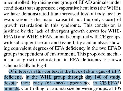
2- The body is a dissipative system; if you remove n-3/n-6, the body changes the way it handles various processes that previously relied on them. It is a mistake in these studies to remove n-6 while keeping the diet essentially the same.
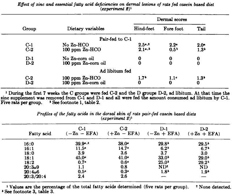
I’ll give an example using zinc, a deficiency of which causes similar skin symptoms:

This table shows an EFAD (HCO) diet and a non-EFAD (corn oil) diet, and both have versions supplemented with zinc (C-2 and D-2); the key point here is that the protein source is casein (this detail will be important later):
Now, in this table, it's the same thing, but supplemented with egg albumin instead of casein:
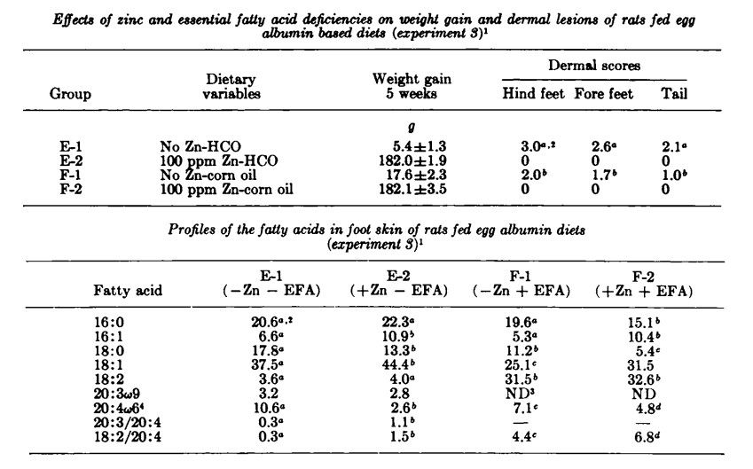
It can be seen from these two tables that EFAD causes skin problems if:
- There is a zinc deficiency
- The proteins are casein-based, rather than egg albumin.
I was curious as to why the type of protein would influence the symptoms, so I remembered this:
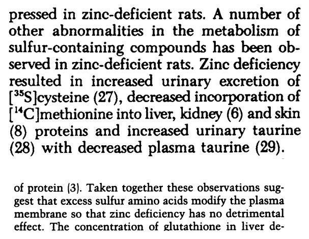
Egg albumin is rich in sulfur-containing amino acids.
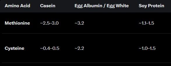
I considered the possibility of contamination with PUFA, but they also use soy protein, which I think theoretically poses a greater risk:
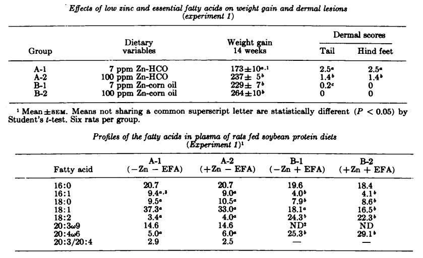
Is it possible that, with enough zinc, they could live indefinitely on an EFAD diet? Who knows... Well, if the problem is humidity, people who live by the beach can easily follow a diet like this and reap the benefits of the EFAD (resistance to endotoxins, inflammation, autoimmune diseases, snake venom, etc.).
And btw, it is possible for a human to live well in EFAD, without skin problems, in a city as well.
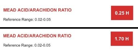
(not mine)Thanks to learnmyhelpless at X for reminding me of zinc and EFAD relationship haha
-
 M Mauritio referenced this topic on
-
Potential Role of the Skin in Hypertension Risk Through Water Conservation
Abstract
Previous basic and clinical investigations have identified various pathogenic factors and determinants of risk that contribute to hypertension. Nevertheless, the pathogenesis of hypertension has not been fully elucidated. Moreover, despite the availability of antihypertensive medications for the management of blood pressure, treatments that address the full spectrum of the pathophysiological defects underpinning hypertension remain to be identified. To further investigate the mechanisms of primary hypertension, it is imperative to consider novel potential aspects, such as fluid management by the skin, in addition to the conventional risk factors. There is a close association between body fluid regulation and blood pressure, and the kidney, which, as the principal organ responsible for body fluid homeostasis, is the primary target for research in the field of hypertension. In addition, the skin functions as a biological barrier, potentially contributing to body fluid regulation. In this review, we propose the hypothesis that changes in skin water conservation are associated with hypertension risk based on recent findings. Further studies are required to clarify whether this novel hypothesis is limited to specific hypertension or applies to physiological blood pressure regulation.
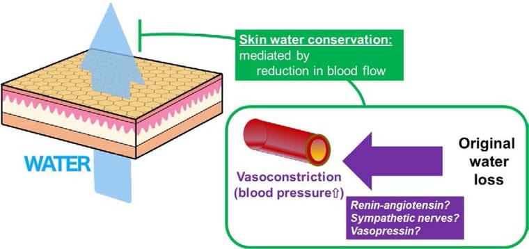
Figure. Fluid loss, skin water conservation, and high blood pressure. Fluid loss activates the systemic water conservation system, leading to high blood pressure, because it is coupled with vasoconstriction. Reduction in skin blood flow contributes to water conservation by suppressing transepidermal water loss.
https://www.ahajournals.org/doi/10.1161/HYPERTENSIONAHA.123.20700Transepidermal water loss and dermal water content for quantitative assessment of cardiac congestion
Congestion assessment is essential for managing congestive heart failure (CHF). Perfusion and tissue water-related skin biophysical properties, namely transepidermal water loss (TEWL) and tissue dielectric constant (TDC), could reflect helpful information regarding circulatory change and oedema severity in these patients. This study compared these biophysical properties between 51 adult participants with CHF and 49 age- and sex-match controls without CHF and assessed their correlations with clinical indicators of congestion. TEWL, epidermal and dermal TDC were measured from the medial and lateral sides of the shin and peri-malleolar areas in a sitting position. Covariate-adjusted multiple linear regressions were used to determine the associations between the biophysical parameters and outcomes. Compared to controls, higher dermal TDC at the shin and medial malleolus, higher epidermal TDC and lower TEWL of the medial shin were observed among CHF individuals and associated with the CHF status regardless of oedema severity, age, sex, and metabolic comorbidities. Medial shin TDC estimates demonstrated the strongest association with CHF status (coefficient [95%CI]: 4.87 [2.06, 7.68]), and positional correlations with other CHF-associated biophysical parameters except for the medial shin TEWL. Subgroup analyses among CHF patients showed positive correlations between the medial shin TDC estimates and the clinical severity of pitting oedema (coefficient [95%CI]: 3.89 [1.11, 6.67]), and the medial shin TEWL estimates and symptomatic CHF status (coefficient [95%CI]: 2.12 [0.13, 4.11]). Given these findings, medial shin TEWL and dermal TDC could be utilised as objective assessments of congestion severity in CHF patients.
https://www.jidonline.org/article/S0022-202X(24)00666-3/fulltextAssessing Vascular Tone and Fluid Balance in Septic and Cardiogenic Shock: A Feasibility Study on Skin Water Loss as a Diagnostic Tool
Background/Objectives: Fluid management in shock remains a clinical challenge, with ongoing debate about optimal guidance. Despite advanced technologies, fluid balance assessment is often inadequate. The SkInShock study investigated whether transepidermal water loss (TEWL) measurements could improve fluid balance estimation and serve as a non-invasive marker of vascular tone in patients with septic or cardiogenic shock. Methods: In this prospective single-center feasibility study (DRKS00027981), TEWL was measured daily in eight mechanically ventilated patients using a Tewameter
(Courage+Khazaka, Cologne, Germany), which quantifies transcutaneous water evaporation. Total daily skin water loss was calculated either via direct TEWL measurements or an estimation formula (6 mL/kg/day + 20%/°C deviation from 37 °C). Systemic vascular resistance index (SVRI) was measured simultaneously using PiCCO technology (Pulsion Medical Systems, Munich, Germany) to evaluate the relationship between TEWL and vascular tone. Results: TEWL values were consistent across most body sites, except the forehead. TEWL-based estimates of skin water loss were significantly lower than formula-based estimates (p < 0.01). Formula-based values overestimated water loss at low TEWL levels and underestimated it at higher levels, with deviations reaching ±100%. While absolute TEWL values did not correlate with SVRI, intra-individually normalized values showed a significant negative correlation, indicating that higher skin water loss corresponded to lower vascular tone. Conclusions: TEWL measurement is feasible in ICU patients and may enhance fluid balance assessment and vascular tone monitoring. Our preliminary findings indicate that this non-invasive method could complement current diagnostics but warrants further investigation in larger cohorts.
https://pmc.ncbi.nlm.nih.gov/articles/PMC12650400/Transepidermal water loss (TEWL): Environment and pollution—A systematic review
Abstract
Introduction
Transepidermal water loss (TEWL) is an objective measurement of skin integrity measured as the amount of water lost across the stratum corneum. TEWL varies greatly across variables such as age and anatomic location, and disruptions in the skin barrier have been linked to inflammatory dermatoses such as psoriasis and atopic dermatitis. Impact of environmental conditions and pollution on TEWL has yet to be determined. Accordingly, this review summarizes effects of environmental conditions and pollution on TEWL.Conclusion
Exposure to pollution is reported to cause increases in TEWL, likely through free radical damage. Significant discrepancies exist among current literature as to the effects of season and climate on TEWL. There is a need to continue examining environmental variables other than temperature and relative humidity, such as atmospheric and steam pressure, that may impact TEWL.
https://pmc.ncbi.nlm.nih.gov/articles/PMC9168018/Dietary fish-oil supplementation in humans reduces UVB-erythemal sensitivity but increases epidermal lipid peroxidation
Abstract
Ultraviolet radiation (UVR)-induced erythema may be mediated in part by free radical-generated tissue damage, including lipid peroxidation. We have examined the effect of dietary fish oil rich in omega-3 fatty acids upon susceptibility to UVB-induced erythema and epidermal lipid peroxidation. Fifteen volunteers took 10 g fish oil, containing 18% eicosapentaenoic acid and 12% docosahexaenoic acid, daily for 3 or 6 months. Sensitivity to UVB was assessed at intervals on fish oil, and 2.5 months after stopping treatment. Paired skin shave biopsies were taken from six subjects, at baseline and 3 months, from both irradiated and control skin. Fatty acid composition was analyzed and thiobarbituric acid-reactive substances measured as an index of lipid peroxidation. With increasing time on fish oil the minimal erythema dose rose progressively, from 18.9 +/- 13.9 mJ/cm2 (mean +/- SD) at baseline to 41.1 +/- 16.6 mJ/cm2 at 6 months, p < 0.01. Ten weeks after stopping fish oil the minimal erythema dose fell to 23.1 +/- 4.9 mJ/cm2, p < 0.05. Epidermal total omega-3 fatty acids rose from 1.8 +/- 0.4% total fatty acids (mean +/- SEM) to 24.2 +/- 3.9% at 3 months, p < 0.01. This was accompanied by a rise in thiobarbituric acid-reactive substances in irradiated skin from 6 +/- 0.3 (mean +/- SEM) to 18.5 +/- 2.6 A532/g skin, p < 0.01. Hence dietary omega-3 fatty acids produce a pronounced reduction in UVB-erythemal sensitivity, although susceptibility of skin to lipid peroxidation is increased. Thus, omega-3 fatty acids may act as an oxidizable buffer, protecting more vital structures from free radical damage.
https://pubmed.ncbi.nlm.nih.gov/8040603/Clinical effects of undershirts coated with borage oil on children with atopic dermatitis: a double-blind, placebo-controlled clinical trial
Abstract
It has been reported that gamma-linolenic acid contained in borage oil is effective against atopic dermatitis. The clinical effects of undershirts coated with borage oil rich in gamma-linolenic acid on atopic dermatitis were evaluated. Thirty-two children, aged 1-10 years, were involved in the clinical control study. Sixteen had worn undershirts coated with borage oil everyday for 2 weeks, and 16 had worn non-coated undershirts as a placebo. Their symptoms were assessed on a 4-point scale. Those children who had worn undershirts coated with borage oil for 2 weeks showed improvements in their erythema and itch, which were statistically significant. Transepidermal water loss from the back was decreased. In the placebo group, there were no statistically significant differences. The undershirts coated with borage oil were found to be statistically effective, and had no side-effects on children with mild atopic dermatitis.
https://pubmed.ncbi.nlm.nih.gov/18078406/The protective effects of Gamma-linolenic acid against indomethacin-induced gastric ulcer in rats
Abstract
The primary goal of the investigation was to analyse the anti-inflammatory and antioxidant properties of Gamma-linolenic acid (GLA) on rats with indomethacin (IND)-induced gastric ulcers. Thirty rats were divided into five groups: Control, IND (50 mg/kg, p.o.), IND pretreated with GLA 100 mg/kg (p.o. for 14 d), IND pretreated with GLA 150 mg/kg (p.o. for 14 d) and IND pretreated with omeprazole (20 mg/kg, p.o. for 14 d). The stomach tissues were examined to calculate the ulcer index and pH and analyse biochemical markers (prostaglandin E2 (PGE2), cyclooxygenase 1 (COX1), TNF-1, IL-6 and intercellular adhesion molecule-1 (ICAM1)) and oxidative stress parameters (malondialdehyde: (MDA), superoxide dismutase (SOD), glutathione (GSH) and CAT (catalase)) as well as undergo histopathological assessment. GLA 100 and 150 mg/kg showed a protective effect against IND-induced gastric damage. It reduced levels of COX1, TNF-1, IL-6 and ICAM and increased PGE2 levels. GLA also normalised antioxidant function by modulating MDA, SOD, GSH and CAT. GLA intervention protects against IND-induced gastric ulcers by restoring oxidant/antioxidant balance and reducing inflammation.
https://www.cambridge.org/core/journals/british-journal-of-nutrition/article/protective-effects-of-gammalinolenic-acid-against-indomethacininduced-gastric-ulcer-in-rats/F942CF859FFD1BB838D27FE3AC2A566CLinoleic acid ameliorates intestinal mucosal barrier injury in early weaned pigeon squabs (Columba livia)
Abstract
The study aimed to investigate whether linoleic acid could improve the intestinal barrier function of squabs under weaning stress conditions. Totally 320 7-d-old weaned squabs were randomly divided into four treatment groups, including control group (CON), 0.7% linoleic acid addition group (LA007), 1.4% linoleic acid addition group (LA014) and 2.1% linoleic acid addition group (LA021). At 21 d, eight squabs were randomly selected from each treatment group for sampling and determination. The results showed that adding linoleic acid could improve (P < 0.05) the body weight of weaned squabs, and LA014 had the best effect. With the increase of linoleic acid dosage, villi height and villi area increased linearly or quadratically (P < 0.05), and reached the maximum in LA021 or LA014, respectively. The linoleic acid supplementation could improve the intestinal tight junction of weaned squabs, and the LA014 was the most significant (P < 0.05). With the linoleic acid increasing, the levels of intestinal IL-6 and TNF-α decreased linearly (P < 0.05), while intestinal IL-10 increased quadratically (P < 0.05) and reached the maximum in LA014. Serum endotoxin and diamine oxidase levels decreased linearly (P < 0.05) and reached the lowest level in LA014. The ultrastructure of villi revealed that the length of ileal microvilli in LA014 was significantly increased (P < 0.05) and the microvilli became dense, and the mitochondria in epithelial cells returned to normal state. Further exploring the mechanism of linoleic acid alleviating intestinal injury caused by weaning stress in squabs, it was found that linoleic acid down-regulated (P < 0.05) the relative protein expression of TLR4, MyD88, phosphorylated JNK, and phosphorylated p38, reducing secretion of pro-inflammatory factors IL-6 and TNF-α. This study indicated that linoleic acid could alleviate intestinal barrier injury of early weaned squabs by down-regulating TLR4-MyD88-JNK/p38-IL6/TNF-α pathway.
https://pmc.ncbi.nlm.nih.gov/articles/PMC10195202/ -
@AlphaZance on the EFAD side of this, Ray Peat went back to the Burr rats in his fats and degeneration article. He said later papers showed Burr's diet was short on B vitamins, and "The disease that appeared in Burr's animals could be cured by fat free B-vitamin preparations, or by purified vitamin B6 when it became available." He also mentioned Burr measured the deficient rats metabolizing 50% faster than rats getting linoleic and linolenic acid. That fits what TexugoDoMel said about skin water loss pushing metabolism up. Has anyone seen the humidity experiments done with the B6 fixed?





Hello! It looks like you're interested in this conversation, but you don't have an account yet.
Getting fed up of having to scroll through the same posts each visit? When you register for an account, you'll always come back to exactly where you were before, and choose to be notified of new replies (either via email, or push notification). You'll also be able to save bookmarks and upvote posts to show your appreciation to other community members.
With your input, this post could be even better 💗
Register Login