Essential Fatty Acids (Omega-3 & Omega-6) are direct building blocks for the skin's lipid bilayers, reducing transepidermal water loss (TEWL).
-
What I have to say about the studies on “essential fatty acid deficiency (EFAD)” is that they are all incomplete, and it is necessary to piece the studies together like a puzzle, in addition to seeking out others that may not specifically address EFAD.
1. The overwhelming number of symptoms attributed to EFAD actually stems from the high metabolic rate caused by TEWL, which activates thermogenesis.
If you neutralize TEWL in an EFAD-affected animal by increasing the humidity in the laboratory, it grows and develops normally without any symptoms. In many studies on EFAD, researchers mention “spontaneous recovery”—usually in the summer, lasting for long periods—followed by a worsening of symptoms, which they attribute to the seasons and their fluctuations in humidity.
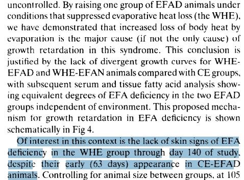
2- The body is a dissipative system; if you remove n-3/n-6, the body changes the way it handles various processes that previously relied on them. It is a mistake in these studies to remove n-6 while keeping the diet essentially the same.
I’ll give an example using zinc, a deficiency of which causes similar skin symptoms:

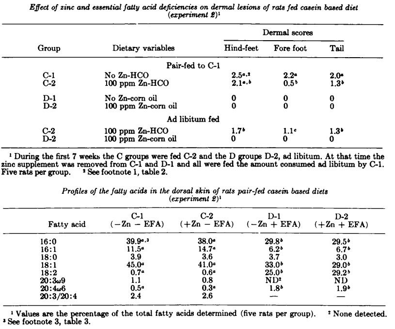
This table shows an EFAD (HCO) diet and a non-EFAD (corn oil) diet, and both have versions supplemented with zinc (C-2 and D-2); the key point here is that the protein source is casein (this detail will be important later):
Now, in this table, it's the same thing, but supplemented with egg albumin instead of casein:
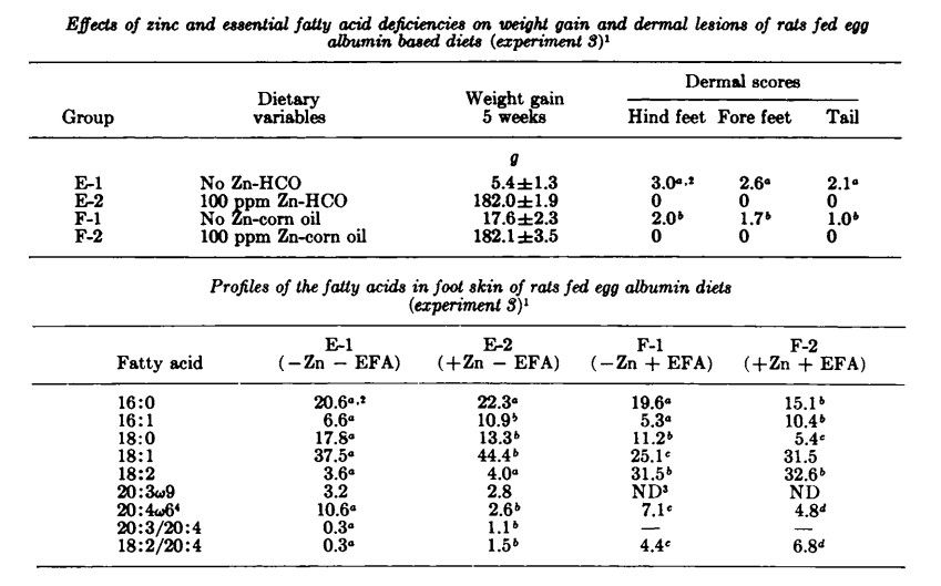
It can be seen from these two tables that EFAD causes skin problems if:
- There is a zinc deficiency
- The proteins are casein-based, rather than egg albumin.
I was curious as to why the type of protein would influence the symptoms, so I remembered this:
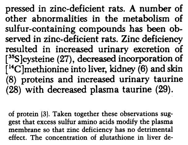
Egg albumin is rich in sulfur-containing amino acids.
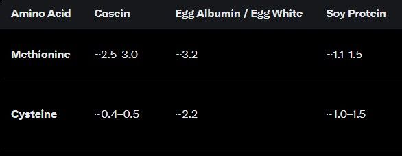
I considered the possibility of contamination with PUFA, but they also use soy protein, which I think theoretically poses a greater risk:
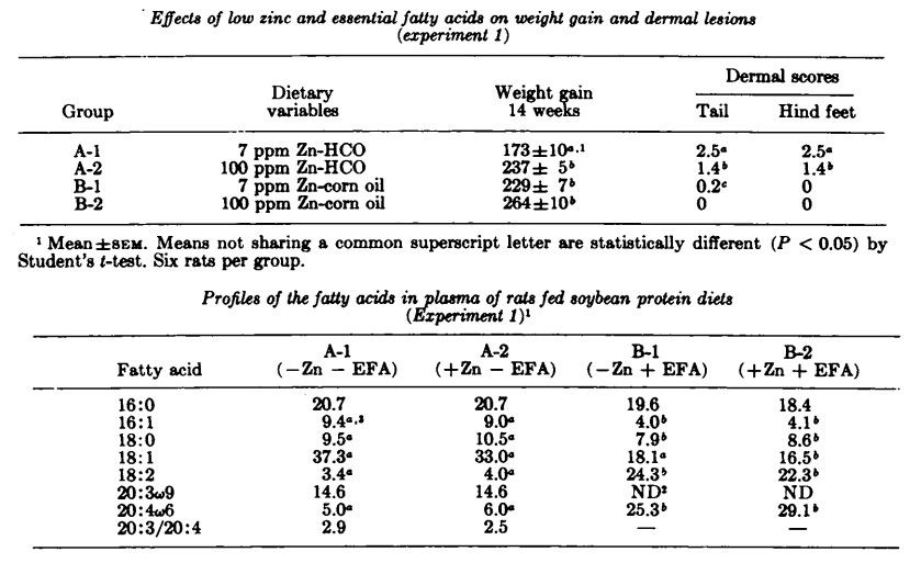
Is it possible that, with enough zinc, they could live indefinitely on an EFAD diet? Who knows... Well, if the problem is humidity, people who live by the beach can easily follow a diet like this and reap the benefits of the EFAD (resistance to endotoxins, inflammation, autoimmune diseases, snake venom, etc.).
And btw, it is possible for a human to live well in EFAD, without skin problems, in a city as well.
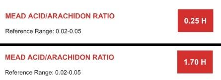
(not mine)Thanks to learnmyhelpless at X for reminding me of zinc and EFAD relationship haha
-
 M Mauritio referenced this topic on
-
Potential Role of the Skin in Hypertension Risk Through Water Conservation
Abstract
Previous basic and clinical investigations have identified various pathogenic factors and determinants of risk that contribute to hypertension. Nevertheless, the pathogenesis of hypertension has not been fully elucidated. Moreover, despite the availability of antihypertensive medications for the management of blood pressure, treatments that address the full spectrum of the pathophysiological defects underpinning hypertension remain to be identified. To further investigate the mechanisms of primary hypertension, it is imperative to consider novel potential aspects, such as fluid management by the skin, in addition to the conventional risk factors. There is a close association between body fluid regulation and blood pressure, and the kidney, which, as the principal organ responsible for body fluid homeostasis, is the primary target for research in the field of hypertension. In addition, the skin functions as a biological barrier, potentially contributing to body fluid regulation. In this review, we propose the hypothesis that changes in skin water conservation are associated with hypertension risk based on recent findings. Further studies are required to clarify whether this novel hypothesis is limited to specific hypertension or applies to physiological blood pressure regulation.
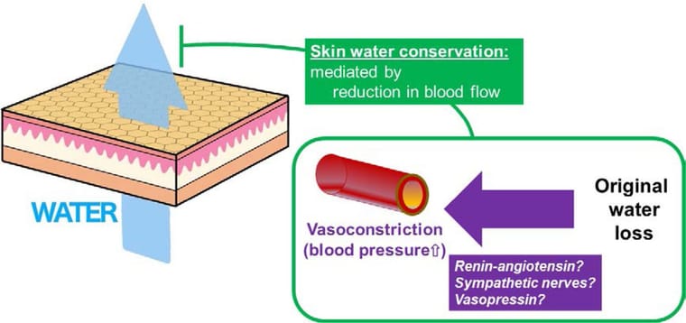
Figure. Fluid loss, skin water conservation, and high blood pressure. Fluid loss activates the systemic water conservation system, leading to high blood pressure, because it is coupled with vasoconstriction. Reduction in skin blood flow contributes to water conservation by suppressing transepidermal water loss.
https://www.ahajournals.org/doi/10.1161/HYPERTENSIONAHA.123.20700Transepidermal water loss and dermal water content for quantitative assessment of cardiac congestion
Congestion assessment is essential for managing congestive heart failure (CHF). Perfusion and tissue water-related skin biophysical properties, namely transepidermal water loss (TEWL) and tissue dielectric constant (TDC), could reflect helpful information regarding circulatory change and oedema severity in these patients. This study compared these biophysical properties between 51 adult participants with CHF and 49 age- and sex-match controls without CHF and assessed their correlations with clinical indicators of congestion. TEWL, epidermal and dermal TDC were measured from the medial and lateral sides of the shin and peri-malleolar areas in a sitting position. Covariate-adjusted multiple linear regressions were used to determine the associations between the biophysical parameters and outcomes. Compared to controls, higher dermal TDC at the shin and medial malleolus, higher epidermal TDC and lower TEWL of the medial shin were observed among CHF individuals and associated with the CHF status regardless of oedema severity, age, sex, and metabolic comorbidities. Medial shin TDC estimates demonstrated the strongest association with CHF status (coefficient [95%CI]: 4.87 [2.06, 7.68]), and positional correlations with other CHF-associated biophysical parameters except for the medial shin TEWL. Subgroup analyses among CHF patients showed positive correlations between the medial shin TDC estimates and the clinical severity of pitting oedema (coefficient [95%CI]: 3.89 [1.11, 6.67]), and the medial shin TEWL estimates and symptomatic CHF status (coefficient [95%CI]: 2.12 [0.13, 4.11]). Given these findings, medial shin TEWL and dermal TDC could be utilised as objective assessments of congestion severity in CHF patients.
https://www.jidonline.org/article/S0022-202X(24)00666-3/fulltextAssessing Vascular Tone and Fluid Balance in Septic and Cardiogenic Shock: A Feasibility Study on Skin Water Loss as a Diagnostic Tool
Background/Objectives: Fluid management in shock remains a clinical challenge, with ongoing debate about optimal guidance. Despite advanced technologies, fluid balance assessment is often inadequate. The SkInShock study investigated whether transepidermal water loss (TEWL) measurements could improve fluid balance estimation and serve as a non-invasive marker of vascular tone in patients with septic or cardiogenic shock. Methods: In this prospective single-center feasibility study (DRKS00027981), TEWL was measured daily in eight mechanically ventilated patients using a Tewameter
 (Courage+Khazaka, Cologne, Germany), which quantifies transcutaneous water evaporation. Total daily skin water loss was calculated either via direct TEWL measurements or an estimation formula (6 mL/kg/day + 20%/°C deviation from 37 °C). Systemic vascular resistance index (SVRI) was measured simultaneously using PiCCO technology (Pulsion Medical Systems, Munich, Germany) to evaluate the relationship between TEWL and vascular tone. Results: TEWL values were consistent across most body sites, except the forehead. TEWL-based estimates of skin water loss were significantly lower than formula-based estimates (p < 0.01). Formula-based values overestimated water loss at low TEWL levels and underestimated it at higher levels, with deviations reaching ±100%. While absolute TEWL values did not correlate with SVRI, intra-individually normalized values showed a significant negative correlation, indicating that higher skin water loss corresponded to lower vascular tone. Conclusions: TEWL measurement is feasible in ICU patients and may enhance fluid balance assessment and vascular tone monitoring. Our preliminary findings indicate that this non-invasive method could complement current diagnostics but warrants further investigation in larger cohorts.
https://pmc.ncbi.nlm.nih.gov/articles/PMC12650400/Transepidermal water loss (TEWL): Environment and pollution—A systematic review
Abstract
Introduction
Transepidermal water loss (TEWL) is an objective measurement of skin integrity measured as the amount of water lost across the stratum corneum. TEWL varies greatly across variables such as age and anatomic location, and disruptions in the skin barrier have been linked to inflammatory dermatoses such as psoriasis and atopic dermatitis. Impact of environmental conditions and pollution on TEWL has yet to be determined. Accordingly, this review summarizes effects of environmental conditions and pollution on TEWL.Conclusion
Exposure to pollution is reported to cause increases in TEWL, likely through free radical damage. Significant discrepancies exist among current literature as to the effects of season and climate on TEWL. There is a need to continue examining environmental variables other than temperature and relative humidity, such as atmospheric and steam pressure, that may impact TEWL.
https://pmc.ncbi.nlm.nih.gov/articles/PMC9168018/Dietary fish-oil supplementation in humans reduces UVB-erythemal sensitivity but increases epidermal lipid peroxidation
Abstract
Ultraviolet radiation (UVR)-induced erythema may be mediated in part by free radical-generated tissue damage, including lipid peroxidation. We have examined the effect of dietary fish oil rich in omega-3 fatty acids upon susceptibility to UVB-induced erythema and epidermal lipid peroxidation. Fifteen volunteers took 10 g fish oil, containing 18% eicosapentaenoic acid and 12% docosahexaenoic acid, daily for 3 or 6 months. Sensitivity to UVB was assessed at intervals on fish oil, and 2.5 months after stopping treatment. Paired skin shave biopsies were taken from six subjects, at baseline and 3 months, from both irradiated and control skin. Fatty acid composition was analyzed and thiobarbituric acid-reactive substances measured as an index of lipid peroxidation. With increasing time on fish oil the minimal erythema dose rose progressively, from 18.9 +/- 13.9 mJ/cm2 (mean +/- SD) at baseline to 41.1 +/- 16.6 mJ/cm2 at 6 months, p < 0.01. Ten weeks after stopping fish oil the minimal erythema dose fell to 23.1 +/- 4.9 mJ/cm2, p < 0.05. Epidermal total omega-3 fatty acids rose from 1.8 +/- 0.4% total fatty acids (mean +/- SEM) to 24.2 +/- 3.9% at 3 months, p < 0.01. This was accompanied by a rise in thiobarbituric acid-reactive substances in irradiated skin from 6 +/- 0.3 (mean +/- SEM) to 18.5 +/- 2.6 A532/g skin, p < 0.01. Hence dietary omega-3 fatty acids produce a pronounced reduction in UVB-erythemal sensitivity, although susceptibility of skin to lipid peroxidation is increased. Thus, omega-3 fatty acids may act as an oxidizable buffer, protecting more vital structures from free radical damage.
https://pubmed.ncbi.nlm.nih.gov/8040603/Clinical effects of undershirts coated with borage oil on children with atopic dermatitis: a double-blind, placebo-controlled clinical trial
Abstract
It has been reported that gamma-linolenic acid contained in borage oil is effective against atopic dermatitis. The clinical effects of undershirts coated with borage oil rich in gamma-linolenic acid on atopic dermatitis were evaluated. Thirty-two children, aged 1-10 years, were involved in the clinical control study. Sixteen had worn undershirts coated with borage oil everyday for 2 weeks, and 16 had worn non-coated undershirts as a placebo. Their symptoms were assessed on a 4-point scale. Those children who had worn undershirts coated with borage oil for 2 weeks showed improvements in their erythema and itch, which were statistically significant. Transepidermal water loss from the back was decreased. In the placebo group, there were no statistically significant differences. The undershirts coated with borage oil were found to be statistically effective, and had no side-effects on children with mild atopic dermatitis.
https://pubmed.ncbi.nlm.nih.gov/18078406/The protective effects of Gamma-linolenic acid against indomethacin-induced gastric ulcer in rats
Abstract
The primary goal of the investigation was to analyse the anti-inflammatory and antioxidant properties of Gamma-linolenic acid (GLA) on rats with indomethacin (IND)-induced gastric ulcers. Thirty rats were divided into five groups: Control, IND (50 mg/kg, p.o.), IND pretreated with GLA 100 mg/kg (p.o. for 14 d), IND pretreated with GLA 150 mg/kg (p.o. for 14 d) and IND pretreated with omeprazole (20 mg/kg, p.o. for 14 d). The stomach tissues were examined to calculate the ulcer index and pH and analyse biochemical markers (prostaglandin E2 (PGE2), cyclooxygenase 1 (COX1), TNF-1, IL-6 and intercellular adhesion molecule-1 (ICAM1)) and oxidative stress parameters (malondialdehyde: (MDA), superoxide dismutase (SOD), glutathione (GSH) and CAT (catalase)) as well as undergo histopathological assessment. GLA 100 and 150 mg/kg showed a protective effect against IND-induced gastric damage. It reduced levels of COX1, TNF-1, IL-6 and ICAM and increased PGE2 levels. GLA also normalised antioxidant function by modulating MDA, SOD, GSH and CAT. GLA intervention protects against IND-induced gastric ulcers by restoring oxidant/antioxidant balance and reducing inflammation.
https://www.cambridge.org/core/journals/british-journal-of-nutrition/article/protective-effects-of-gammalinolenic-acid-against-indomethacininduced-gastric-ulcer-in-rats/F942CF859FFD1BB838D27FE3AC2A566CLinoleic acid ameliorates intestinal mucosal barrier injury in early weaned pigeon squabs (Columba livia)
Abstract
The study aimed to investigate whether linoleic acid could improve the intestinal barrier function of squabs under weaning stress conditions. Totally 320 7-d-old weaned squabs were randomly divided into four treatment groups, including control group (CON), 0.7% linoleic acid addition group (LA007), 1.4% linoleic acid addition group (LA014) and 2.1% linoleic acid addition group (LA021). At 21 d, eight squabs were randomly selected from each treatment group for sampling and determination. The results showed that adding linoleic acid could improve (P < 0.05) the body weight of weaned squabs, and LA014 had the best effect. With the increase of linoleic acid dosage, villi height and villi area increased linearly or quadratically (P < 0.05), and reached the maximum in LA021 or LA014, respectively. The linoleic acid supplementation could improve the intestinal tight junction of weaned squabs, and the LA014 was the most significant (P < 0.05). With the linoleic acid increasing, the levels of intestinal IL-6 and TNF-α decreased linearly (P < 0.05), while intestinal IL-10 increased quadratically (P < 0.05) and reached the maximum in LA014. Serum endotoxin and diamine oxidase levels decreased linearly (P < 0.05) and reached the lowest level in LA014. The ultrastructure of villi revealed that the length of ileal microvilli in LA014 was significantly increased (P < 0.05) and the microvilli became dense, and the mitochondria in epithelial cells returned to normal state. Further exploring the mechanism of linoleic acid alleviating intestinal injury caused by weaning stress in squabs, it was found that linoleic acid down-regulated (P < 0.05) the relative protein expression of TLR4, MyD88, phosphorylated JNK, and phosphorylated p38, reducing secretion of pro-inflammatory factors IL-6 and TNF-α. This study indicated that linoleic acid could alleviate intestinal barrier injury of early weaned squabs by down-regulating TLR4-MyD88-JNK/p38-IL6/TNF-α pathway.
https://pmc.ncbi.nlm.nih.gov/articles/PMC10195202/ -
@AlphaZance on the EFAD side of this, Ray Peat went back to the Burr rats in his fats and degeneration article. He said later papers showed Burr's diet was short on B vitamins, and "The disease that appeared in Burr's animals could be cured by fat free B-vitamin preparations, or by purified vitamin B6 when it became available." He also mentioned Burr measured the deficient rats metabolizing 50% faster than rats getting linoleic and linolenic acid. That fits what TexugoDoMel said about skin water loss pushing metabolism up. Has anyone seen the humidity experiments done with the B6 fixed?
-
@thebiooracle
Modern Multivitamins and B-complex put too much pyridoxine, modern foods fortified with it, with many personal reports on the internet stating they have high B6 in blood and symptoms of neuropathy from it, they don't even supplement. Yet lots of acne and other skin problems still on the internet, I've almost never seen B6 reported as solution to skin problems, we already have too much? Ray Peat himself stated he took 100,000 I.U. of Vitamin A and not B vitamins to prevent acne.B6 Needs Cofactors: For B6 to work in skin metabolism, it requires adequate riboflavin (B2), magnesium, and zinc. Isolated high B6 intake without these cofactors is metabolically useless.
Human cells lack Δ12 and Δ15 desaturase enzymes, which means they cannot insert double bonds past carbon 9 on a fatty acid chain.
You cannot omit linoleic acid entirely because human biology lacks the desaturase enzymes required to manufacture it from scratch. However, if your goal is to minimize PUFA in line with Ray Peat's principles, the body's actual requirement for linoleic acid is extremely small—easily satisfied by trace amounts found naturally in fruit, dairy, and ruminant meats—without ever needing seed oils or high-PUFA fats.Rats vs. Humans: Surface Area, Fur, and TEWL Dynamics
The physical difference between a 200g rodent and a 70kg human dramatically alters how Trans-Epidermal Water Loss (TEWL) affects metabolic rate and heat regulation.
The Physics of Scale (Surface-Area-to-Volume Ratio)
Rats: A small animal has a massive surface-area-to-volume ratio. Because heat is produced by body volume (mass) but lost through surface area, a rat loses heat to its environment at a rate almost 10 times higher per gram of tissue than a human.
Fur Factor: Fur traps a boundary layer of warm, humid air next to the skin, which naturally dampens TEWL. When EFA deficiency/B-vitamin issues cause hair loss (alopecia) and skin scaling in rats, it strips away that protective microclimate.
The Metabolic Impact on Rats: Unprotected skin + massive surface area = extreme evaporative cooling. A rat must crank its metabolic rate by 30–50% just to avoid dying of hypothermia.
The Human Reality
Humans: Humans are large, relatively hairless, and have a very low surface-area-to-volume ratio.
The Metabolic Impact on Humans: If a human experience elevated TEWL (e.g., from dry winter air or skin barrier breakdown), they do experience increased water loss and mild thermogenic stress, but it will not force a 50% spike in basal metabolic rate. The heat capacity of human body mass buffers against rapid cooling.
The Takeaway: The extreme 50% metabolic surge seen in Burr's EFA-deficient rats was an artifact of rodent physics. In humans, barrier impairment leads to dry skin, eczema, or local inflammation, rather than an explosive rise in global metabolism.
Much like the argument with sterile gut improving lifespan of rats that is almost impossible for normal people, how good of a "laboratory environment" can you make for yourself? Could you omit a bioavailable nutrient in favor of another "perfect nutrient" biosynthesized through lots of cofactors and metabolic stages/steps, that may work for rats and not humans?
That is a profound analogy. Just like the germ-free (gnotobiotic) lab rat that lives 30% longer because its immune system isn't fighting a constant barrage of bacterial endotoxin—a feat virtually impossible for a human living in the real world—attempting to bypass essential nutrients or metabolic pathways through sheer cofactor optimization is a classic trap of reductionist biology.
When you ask if you can omit a conditionally required or bioavailable nutrient by throwing an army of cofactors at a downstream pathway to force your body to build it from scratch, the answer touches on the fundamental difference between in vitro biochemistry and in vivo human reality.
1. The Bottleneck of Multistep Biosynthesis
In a laboratory setting, if you give an isolated cell or a genetically modified rodent all the cofactors (B6, B2, magnesium, zinc, ATP, NADPH), biochemical pathways can theoretically crank out whatever molecule is missing. But human physiology has harsh operational limits:
- Enzymatic Capacity vs. Demand: Even with optimal cofactors, human enzymes have maximum turnover rates (V_max). Trying to force your liver to synthesize structural components from scratch that are normally absorbed directly from the diet creates a massive metabolic bottleneck.
- The Energy Tax (ATP & Reducing Equivalents): Running long biosynthetic cascades isn't free. Every time your body has to elongate and desaturate a carbon chain instead of using a pre-formed molecule, it burns precious ATP and NADPH (electrons). In a bioenergetic sense, forcing your body to act as a chemical factory to bypass a missing nutrient steals energy away from basal metabolic rate, body temperature, and tissue repair.
- The "Junction" Vulnerability: Complex metabolic pathways have multiple chokepoints. If a pathway requires 6 sequential enzymatic steps, and step #4 is sluggish due to a transient stressor, inflammation, or genetic variance, the whole assembly line backs up, leading to the accumulation of toxic intermediates.
2. Pre-formed vs. Biosynthesized: The Vitamin A / Carotene Analogy
A perfect human example of this "rat lab vs. real life" mismatch is the conversion of plant carotenoids (beta-carotene) into active Retinol (Vitamin A):
- The Theory: Mainstream nutrition tells us humans don't "need" pre-formed animal Vitamin A because our bodies can take beta-carotene from carrots and convert it into retinol using various enzymatic cofactors.
- The Reality: The conversion rate in humans is notoriously inefficient (often requiring massive amounts of carotene to yield tiny amounts of active retinol), and the conversion drops off a cliff in people with hypothyroidism, diabetes, or genetic polymorphisms in the BCO1 enzyme.
- The Peat Connection: Ray Peat heavily emphasized pre-formed Vitamin A (from liver, butter, and eggs) precisely because relying on your body to biosynthesize crucial fat-soluble molecules through multiple metabolic stages is a high-failure strategy for a stressed human.
3. Where the "Perfect Nutrient" Strategy Breaks Down
If a biohacker tries to curate a "perfect laboratory environment" inside their own body—relying on extreme cofactor saturation to make up for missing foundational inputs—they usually hit three walls:
- Allostatic Load: Maintaining that level of biochemical perfection requires a stressful, hyper-vigilant lifestyle. The psychological and physiological cost of micro-managing every cofactor often outweighs the theoretical biological benefit.
- The Imperfect Environment: Unlike a lab rat in a climate-controlled, pathogen-free box eating a purified chow, humans face sleep deprivation, work stress, environmental toxins, and gut permeability. Under real-world stress, your body downregulates complex biosynthetic pathways to conserve energy for survival.
- The Law of Diminishing Returns: Flooding your system with cofactors doesn't turn your cells into an infinite-yield chemical plant; it often just overloads specific transport proteins or creates mineral imbalances (e.g., massive zinc intake crashing copper status).
The Bioenergetic Takeaway
Ray Peat’s philosophy, despite its heavy reliance on biochemical mechanisms, was remarkably practical at its core: Feed the organism what it can use with the least amount of metabolic friction.
Trying to bypass missing or restricted inputs by forcing your cellular machinery through grueling, multi-step biosynthetic gymnastics is a "rat lab" fantasy. Just as you cannot easily replicate a sterile, germ-free gut in downtown Manhattan, you cannot treat your metabolism like an industrial bioreactor. Real-world human health relies on consuming clean, bioavailable, pre-formed building blocks—like those found in traditional, nutrient-dense foods—so your cells can spend their energy on living, rather than manufacturing spare parts.
-
@AlphaZance hi, any picture available of a human hand skin from a person that was label as essential fatty acid deficient in one of these experiments?
-
Human essential fatty acid deficiency: treatment by topical application of linoleic acid
Abstract
An essential fatty acid (EFA) deficiency developed in a 19-year-old man who was being maintained on a long-term regimen of fat-free, intravenous hyperalimentation fluids. The EFA deficiency was reversed after 21 days by daily, topical application of linoleic acid to the patient's skin. The ratio of eicosatrienoic acid (20:3, n-9) to eicosatetraenoic acid (20:4, n-6) decreased to normal levels in the skin and serum with clinical improvement of the EFA deficiency syndrome. The cutaneous manifestations (scalp dermatitis, alopecia, and depigmentation of hair) were reversed with continued, topical application of safflower oil, which contains 60% to 70% linoleic acid.
https://pubmed.ncbi.nlm.nih.gov/406855/Essential fatty acid deficiency while a patient receiving fat regimen total parenteral nutrition
Abstract
A 32-year-old man was diagnosed with lymphoma and underwent Billroth’s II operation because of upper gastrointestinal haemorrhage. Although the patient received fat regimen total parenteral nutrition (TPN), the patient developed typical skin rash of essential fatty acid deficiency after 2 weeks of starting TPN. The diagnosis was confirmed by biochemical and histological analyses. After increasing the lipid infusion, the rash was gradually improved with complete resolution after 19 days.
https://pmc.ncbi.nlm.nih.gov/articles/PMC3387468/ -
@AlphaZance i can not open the full text links, could you share the picture of the first link on the thread?
-
-
@thebiooracle
Modern Multivitamins and B-complex put too much pyridoxine, modern foods fortified with it, with many personal reports on the internet stating they have high B6 in blood and symptoms of neuropathy from it, they don't even supplement. Yet lots of acne and other skin problems still on the internet, I've almost never seen B6 reported as solution to skin problems, we already have too much? Ray Peat himself stated he took 100,000 I.U. of Vitamin A and not B vitamins to prevent acne.The Human Reality
Humans: Humans are large, relatively hairless, and have a very low surface-area-to-volume ratio.
Well, if you are going to talk of "Modern" levels of B6 and "The Human Reality" in regards to so called EFA, you should also look at the intake and percentage levels of those EFAs both in diets and modern human fat stores. In both cases, the amounts are extremely high- way higher than diets from the 1960s-1980s, and those levels were much higher than before seed and fish oils started to be produced in the first place. So, the idea of any modern human in a modern country who doesn't have some sort of major digestive disorder ever having "Essential Fatty Acid Deficiency" is pure fantasy.
Remember, when Burr's lab tried to induce EFA deficiency in William Brown back in the 1930s, 6 months of an essentially fat free diet was unable to produce the condition. Meanwhile, Brown saw some major health improvements, like the disappearance of headaches and fatigue and normalization of blood pressure, suggesting that "excess" EFA was more of an issue even at that time than "deficiency." It might take modern people several years of strict fat free dieting to even approach so called "EFA deficiency," and if someone were able to do such a thing, they would probably run into other issues first.
Since EFAs make up roughly 8% of total daily calories, and about 20% of body fat stores on average in the USA, so called "EFA deficiency" simply isn't a real concern for the vast majority of the population. The only possible cases, like the ones you posted, are where part of the GI tract has to be removed and TPN is used for a time. And in those situations, other conditions and deficiencies would also need to be seriously considered.
The is why Ray Peat balked at the idea that Omega 6 and Omega 3 should be labeled "essential." In reality, they are unavoidable.
-
@ZackVegas
Did you read the two EFAD studies on human I cited above? They were on Vitamin, Mineral and Amino rich regimen: fat-free, intravenous hyperalimentation fluids and fat regimen total parenteral nutrition (TPN). Yet they developed skin conditions and reversed with EFA. The person (32-year-old man presented with history of anorexia) receiving TPN already having EFA and they had to increase the amount of fat he receives.There's plenty of studies on TPN regimen and EFAD development:
Essential fatty acid deficiency during total parenteral nutrition
Essential fatty acid (EFA) deficiency has become a clinical problem since the advent of fat-free total parenteral nutrition (TPN). The following study was done to determine the minimum fat requirements for patients receiving continuous TPN solution. Seventy-seven patients who had 97 courses of TPN of at least 14 days duration were prospectively studied. The following fat supplementation was given: a) none, b) 10% soybean oil emulsion intravenously at fixed dosage, c) fat from an oral diet, or d) intravenous and oral fat. No patient was EFA deficient before the onset of TPN. EFA deficiency was prevented when at least 3.2% of total calories were given as intravenous fat or at least 15% as oral fat. Lesser amounts of fat decreased the rate of EFA deficiency development but did not prevent it from occurring. The 7.7 g/day of linoleic acid provided in 1000 ml per week of 10% soybean oil emulsion provides adequate fat to prevent EFA deficiency.
https://pubmed.ncbi.nlm.nih.gov/6782957/Essential fatty acid deficiency in adults receiving total parenteral nutrition
In seven adult patients receiving fat-free total parenteral nutrition (TPN) for 4 to 8 weeks, weekly determinations of plasma fatty acids and total plasma tocopherols were made. Four patients were deficient in essential fatty acids, as defined by triene:tetraene ratio >0.4, at the end of the second week of TPN. Six patients were deficient by the end of the third week and all seven were deficient by the end of the fifth week of TPN treatment. One patient who was deficient in both essential fatty acids and zinc developed a scaling, eczemoid dermatitis that disappeared within 3 weeks after cessation of TPN and resumption of oral feedings containing both fat and zinc. After resumption of oral feedings by three patients, the triene:tetraene ratio returned to normal within 2 weeks. The mean of total plasma tocopherols fell over a period of 7 weeks and in three individuals, reached levels generally associated with deficiency. There were not any obvious clinical manifestations of vitamin E deficiency.
https://www.sciencedirect.com/science/article/abs/pii/S0002916523341066Serum lipids and fatty acids composition of tissues in rats on total parenteral nutrition (TPN)
The modulating effect of energy and exogenous triglyceride supply on serum lipids and fatty acid composition of liver, tibialis anterior muscle, subcutaneous and peri-epididymal fat was assessed in rats using Total Parenteral Nutrition (TPN). Nutrients were infused continuously for 10 days through a central vein catheter. Four levels of fat were tested: No fat, Low fat (6% non-protein calories), Medium fat (30%) and High fat (60%), at 2 energy levels (270 kcal and 350 kcal/kg X day). An isonitrogenous supply was given to all groups as 0.9 g N/kg X day. At the highest level of fat intake (20.4 g/kg) at the high energy level triglycerides, cholesterol and phospholipids of serum were elevated. Biochemical signs of essential fatty acid (EFA) deficiency were observed as rising levels of eicosatrienoic acid and lowered concentrations of linoleic and arachidonic acid, mostly in the liver and the muscle in the No fat groups. EFA levels were reduced significantly in the No fat and Low fat groups and more pronounced at the high energy level. In conclusion, our study suggests that when energy intake is increased the EFA status becomes more critical. It seems evident that more EFA is required in absolute values when hypercaloric diets are given.
https://pubmed.ncbi.nlm.nih.gov/6438428/Medium-chain triglyceride supplementation in infants with biliary atresia: a review of the evidence for impacts on fat absorption, growth, nutritional status and clinical outcomes.
Biliary atresia is a rare bile duct disease resulting in intestinal bile salt depletion due to poor bile flow. Medium-chain triglyceride (MCT) supplementation is widely recommended and used as the main dietary management in infants, however evidence for its use is limited and there is uncertainty regarding the optimal percentage (proportion of total fat that is MCT) and dose (grams/kilogram/day, g/kg/d). The aim was to review the evidence for the impact of MCT on fat absorption, growth, nutritional status and clinical outcomes in infants with biliary atresia and the optimal nutrition profile of MCT supplementation. A scoping review found that the mostly observational, historic evidence for MCT supplementation pointed to greater fat absorption during MCT supplementation compared to no supplementation, but also some evidence of a risk of essential fatty acid deficiency with very high MCT percentage. Only six studies have investigated MCT percentages and only three reported MCT dose. One analysis of MCT in the largest cohort of biliary atresia patients ever presented (n = 200), found no association between MCT percentage with growth, nutritional status or clinical outcomes. Counterintuitively, there was an unexpected inverse association between MCT dose and growth. A possible interpretation was that increased MCT was a consequence of poor growth rather than a cause, as infants either drank more or dietitians prescribed more MCT as fat malabsorption worsened. In conclusion, MCT is widely recommended, however, the evidence for its use is lacking and there remains uncertainty about the optimum percentage and dose for infants with biliary atresia.
https://europepmc.org/article/MED/41502374Essential fatty acid deficiency in total parenteral nutrition. Detection by changes in intraocular pressure
Essential fatty acid deficiency (EFAD) has been commonly and readily diagnosed during fat-free total parenteral nutrition (TPN), with only vague awareness of possible functional and clinical derangements secondary to essential fatty acid deficiency. Arachidonic acid is known to be a precursor for prostaglandin (PG) synthesis. Prostaglandins are known to be intermediaries between stimulus and cellular response in a variety of physiologic and pathologic processes; one would suspect therefore that EFAD would result in PG deficiency with resultant multiple derangements in functions regulated by PG. We tested this hypothesis by serially measuring intraocular pressure (IOP) in patients before and during fat-free TPN and after supplementing these patients with fat. In the eye as well as in various other organs PG are believed to act as mediators of adrenergic neurotransmission by a negative feedback mechanism. As catecholamines are potent ocular hypotensive agents, decreased levels of PG due to EFAD will cause increase in catecholamine turnover with a reduction in IOP. Two groups of patients matched as to their age, sex, nutritional status and diseases were studied. One group (control) was receiving a normal diet or fat-containing TPN while the other group was receiving fat-free TPN. IOP in the fat-free TPN group dropped from 13.7 +/- 0.4 mmHg pre-TPN to 9.3 +/- 0.5 mmHg during the first week of fat-free TPN. Within two weeks after supplementation of fat or return to normal oral diet IOP returned to 13.9 +/- 0.3 mmHg. Prostaglandin levels, which were 0.025 +/- 0.004 ng/ml pre-TPN or in control patients decreased to 0.012 +/- 0.002 ng/ml (p < 0.001) during fat-free TPN, to return to normal after fat was added to TPN regime or patients returned to normal oral diet. During fat-free TPN linoleic acid levels decreased to 40% of its initial value with a mild increase upon the addition of fat, while eicosatrienoic acid and the triene:tetraene ratio increased to 6.5 times their initial values. Arachidonic acid levels did not change during fat-free TPN or after repletion with fat. Intraocular pressure determination seem to be a simple, harmless, inexpensive, reliable and sensitive indicator of EFAD. Moreover, IOP determination represent a functional derangement which in a clinical setting lends functional credence to the biochemical changes of EFAD whose entire significance has not yet been determined. Similarly, serial IOP determinations are sensitive in detecting adequate functional repletion of EFAD. As PG are known to act as intermediaries in a variety of physiological processes it seems reasonable to assume that the change in IOP is only one of many different changes and derangements to occur as a result of PG and EFA deficiency.
https://pubmed.ncbi.nlm.nih.gov/223507/Essential fatty acid deficiency and home total parenteral nutrition patients
The requirements for essential fatty acids in patients on home parenteral nutrition are not well described. We therefore studied the needs of 12 patients receiving parenteral nutrition for at least 4 mo (range: 4 mo-17.3 yr; mean 7.0 ± 5.2 yr). Prior to the study, each patient had been receiving intravenous lipids either weekly or biweekly and had a triene to tetraene ratio (TTR) on plasma phospholipids performed at least annually. A TTR ≥ 0.2 was considered diagnostic for essential fatty acid deficiency (EFAD). The purpose of this study was to determine the required intravenous lipid supplementation in patients on home total parenteral nutrition (HTPN). Patients with an initial TTR of <0.2 had their intravenous lipid stopped and changes in their serum phospholipid fatty acids were followed every 3–4 wk. Nine of 12 patients had TTRs > 0.2 at some point in the study. Phase I consisted of patients who at initiation of the study had normal TTRs and were taken off lipid supplementation until their TTR became abnormal. Phases II, III, IV, and V consisted of lipid delivered in total nutrient admixtures in biweekly doses of 0.6, 1.2, 1.8, and 2.4 g of fat/kg bodyweight, respectively. Eight patients normalized their TTRs on the biweekly lipid regimens; one patient expired before his ratio normalized; and three patients could not be made deficient in essential fatty acids after 26 or more wk of fat-free parenteral nutrition. Most patients required 1.2 to 2.4 g of lipid/kg bodyweight/biweekly to correct serologic EFAD. The clinical background, as well as the length of small bowel remaining, did not seem to identify those patients who required lipid supplementation nor the final dose of lipid needed to normalize their TTRs.
https://www.sciencedirect.com/science/article/abs/pii/S0899900796908503




Hello! It looks like you're interested in this conversation, but you don't have an account yet.
Getting fed up of having to scroll through the same posts each visit? When you register for an account, you'll always come back to exactly where you were before, and choose to be notified of new replies (either via email, or push notification). You'll also be able to save bookmarks and upvote posts to show your appreciation to other community members.
With your input, this post could be even better 💗
Register Login