Placing this video in the art directory wouldn't do justice to it, as it transcends art.
Posts made by Amazoniac
-
RE: Humorous musingsposted in The Junkyard
-
Sunlight and its effectsposted in Literature Review
"The mechanisms driving local and systemic UV immune suppression are different. Although much is known about how UV suppresses local cutaneous immune responses, the mechanisms by which UV suppresses distant immunity are only just being revealed. Our study has shown for the first time, to our knowledge, that UV exposure of the skin systemically alters T cell recirculation, preferentially trapping naive and, to a lesser extent, central memory T cells in the UV-exposed skin-draining lymph nodes. This cellular sequestration away from peripheral blood was the result of a UV-triggered increase in SphK activity, leading to elevated levels of S1P in regional lymph nodes. This increase in S1P in turn caused a downregulation of S1P1 expression on T cells leading to their sequestration."
"Pharmacological interference with normal lymphocyte trafficking is a highly effective way to induce therapeutic immune suppression. Natalizumab for example, is an mAb that prevents leukocyte trans-migration by blocking α4β1 integrin leukocyte adhesion to endothelial VCAM-1 (54). Another example is fingolimod, a functional S1P receptor agonist that induces the internalization and downregulation of S1P1, the functional result of which is a reduction in circulating leukocytes (35), in particular naive and central memory T cells (34), and an accumulation of T cells within lymph nodes (36). Interference with normal lymphocyte trafficking following administration of natalizumab and fingolimod causes profound systemic immune suppression that is effective in the treatment of autoimmune diseases, particularly MS (55, 56). We have shown in this study that exposure to UV, which is a known immune suppressant and associated with protection from MS (4), also interferes with normal lymphocyte trafficking."
"The ability of UV to alter T cell recirculation, promote the expansion of UV-activated Tregs (18), and inhibit T cell proliferation upon stimulation (16) provides a potent formula for the comprehensive suppression of cell-mediated immunity. The trapping of naive T cells in the skin-draining lymph nodes maximizes the chance of interactions with other cellular mediators of UV-induced immune suppression, such as dendritic cells (63), mast cells (37), B cells (50, 64), and UV-Tregs (19). It is known that if an Ag is presented during the first 24–72 h of exposure to UV, T cell activation is suppressed (16) in part by UV-Treg and B cell production of IL-10 (19, 20, 65). We have now shown that even if activation of naive T cells occurs, UV not only limits clonal expansion (16), but it also interferes with normal lymphocyte recirculation by reducing the likelihood of cells exiting the lymph nodes."
-
RE: Conditional problems with vitamin A: a place for sane discussionsposted in Literature Review
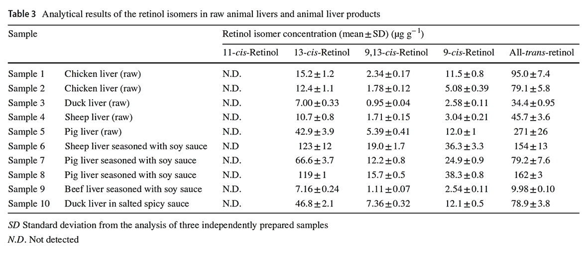
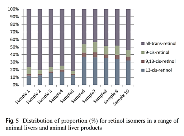
"Considering that the biological activities of isomers are lower than all-trans-retinol, the knowledge of retinol isomer profile in liver is useful for a precise estimation of safe dietary intake levels of animal liver."
"[..]when the determination can not separate retinol isomers, total retinol activity in animal liver products may be overestimated."
-
Interactions between vitamins A and Dposted in Literature Review
To start a discussion on these two toxins..
"It has been demonstrated that TR and RAR interact in solution not only with RXR but also with VDR (35–37). Therefore, it was possible that formation of inactive TR-VDR and RAR-VDR heterodimers which either do not bind or bind in a transcriptionally inactive form to the T3 and RA response elements could contribute to the transcriptional repression observed in pituitary cells. We were not able to find TR-VDR or RAR-VDR binding to the palindromic response element, showing a low affinity, if any, of these heterodimers for the element. However, VDR very effectively inhibited binding of TR-RXR and RAR-RXR by formation of VDR-RXR heterodimers which bind with a much lower affinity than does either TR-RXR or RAR-RXR to the TRE(PAL). More interestingly, VDR also competed TR monomeric or homodimeric binding to this element, demonstrating that VDR sequesters TR (and most likely also RAR) and prevents binding to the element and transcriptional activation."
"Schräder et al. (36) have reported that TR-VDR heterodimers bind almost as strongly as VDR-RXR heterodimers to the osteopontin VDRE. In contrast, we did not detect significant TR-VDR or RAR-VDR binding to this element. Also, with this VDRE we found that TR and RAR displaced not only VDR-RXR binding but also VDR homodimeric binding. Taken together, our data suggest the formation of TR-VDR and RAR-VDR heterodimers which have low affinities by either the TRE or the VDRE and can inhibit binding of more active heterodimers with RXR by competition."
-
Pregnane X receptor and the fat-soluble vitaminsposted in Literature Review
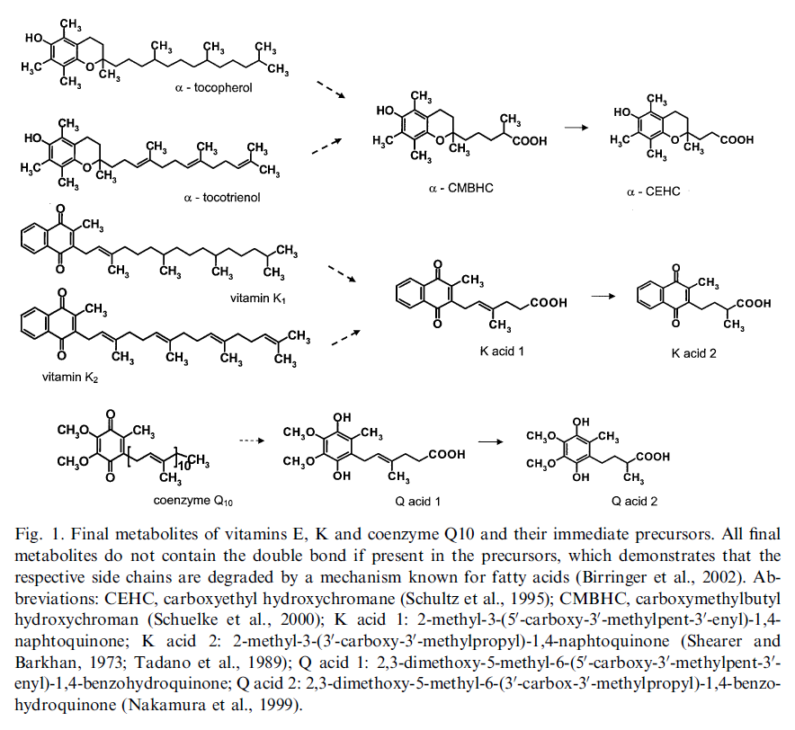
Homologous metabolic and gene activating routes for vitamins E and K
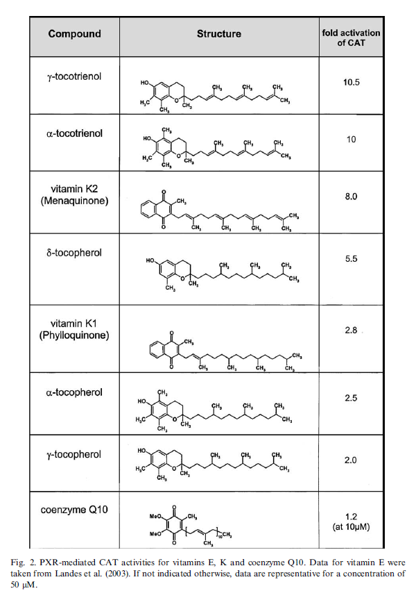
"Vitamins E and K share structurally related side chains and are degraded to similar final products. For vitamin E the mechanism has been elucidated as initial w-hydroxylation and subsequent b-oxidation. For vitamin K the same mechanism can be suggested analogously. w-Hydroxylation of vitamin E is catalyzed by cytochrome P450 enzymes, which often are induced by their substrates themselves via the activation of the nuclear receptor PXR. Vitamin E is able to induce CYP3A-forms and to activate a PXR-driven reporter gene. It is shown here that K-type vitamins are also able to activate PXR. A ranking showed that compounds with an unsaturated side chain were most effective, as are tocotrienols and menaquinone-4 (vitamin K2), which activated the reporter gene 8–10-fold. Vitamers with a saturated side chain, like tocopherols and phylloquinone were less active (2–5-fold activation). From the fact that CYPs commonly responsible for the elimination of xenobiotics are involved in the metabolism of fat-soluble vitamins and the ability of the vitamins to activate PXR it can be concluded that supranutritional amounts of these vitamins might be considered as foreign."
↳ Vitamin E activates gene expression via the pregnane X receptor
-
RE: New "Mission" of RPFposted in Bioenergetics Discussion
I wasn't aware of the mirrored behavior, but it's indeed preferable for members to assume that the characteristic authoritarianism was disinhibited after an ideological justification was found (it happened to be poison A, but could be anything else) rather than being a product of some sinister conspiracy. The pattern of conduct remains the same, only amplified now because of overconfidence to unleash it.
The recent disposals aren't much different in quality than the elimination of members who disagreed with Ray on dietary minutiae in the past, dumping along without remorse whoever questioned.
On the last month, probably more than 50 members were eliminated over nothing, but we never know the full extent of the mess because many moves are sneaky and some members disappear afterwards. For example, what if CreakyJoints or Mauritio didn't voice?
By the way, to ask why a peer was banned isn't back-seat moderation. To seek an explanation not only is forbidden, but treated with punishment. What a joke.
The Ray Peat Forum disappointed the majority of the community formed through Ray, not realizing that those who remained supporters, indifferent to the absurds, may ditch them at any time to be embraced by the groups run by the very proponents that the staff promote. Professor Garrett has been a parasite of the forum since its creation, but it seems that its leaders are fully committed to reversing roles.
Reliance on social media posts by charlatans to keep the platform active is risky because supporters may soon realize that they can interact elsewhere without having to pay questionable fees, on existing groups dedicated to the ideology being imposed on the forum, while still consulting the material there. Giveaways are entertaining, but for $77/year, they may buy 3 products of those without depending on luck.
Since it's not lucrative to buy products from your own store, at this elimination pace, views count may become an important metric and putting barriers that lower them can be counterproductive.
On one hand, it's fair for the lion to not want the content to be scraped because of the maintenance factor throughout time. On the other hand, it's a collective effort that he's trying to appropriate.
That he panicked at the possibility of no longer possessing the material shows the degree of leadership insecurity. Otherwise, people could copy the information as much as they wanted, but they wouldn't be able to recreate the forum experience elsewhere because the leaders are playing a fundamental role, are adored and irreplaceable.
-
RE: Resources for authorsposted in Bioenergetic Development
I gathered them little by little and the collection was available in a private group where I communicate with my gurus.
The English is better now because of conscious effort to moderate the Prolactinese and keep the speech minimally understandable. However, I continue to reject most of the formalism in everyday writing, for treating it as a conversation and to not depersonalize it too much in trying to eliminate mannerisms. As an example, overuse of adverbs is condemned by writers, but I insist for that rebel quota. I don't bother with correctors for informal writing.
This article relates to the second part of your post. But being on the passenger's seat is different than being the driver, so I don't think that reading a lot will be much effective in improving writing skills, although it helps (enhancing vocabulary would be an example). In support of the need of practice, try to learn a new language by only receiving information and applying it once you reach a satisfactory level: it's likely that you'll struggle.
-
RE: Mike Fave is on fireposted in Bioenergetics Discussion
@Peatful said in Mike Fave is on fire:
@Amazoniac
Curious
Do you know him personally?
And / Or
Have you consulted with him for health reasons?If I'm alive, it means that I haven't consulted with him.Humor aside, the post was not paid promotion.
We have communicated before, but we're not close.
In case the tags seemed sketchy, people are often looking for assistance to tackle issues while preferring for the helper to be familiar with Ray's work. Our views differ on occasion, but Miguel is someone within the community that I would recommend to others.
-
RE: Conditional problems with vitamin A: a place for sane discussionsposted in Literature Review
Gut commensals expand vitamin A metabolic capacity of the mammalian host
"Here, using liquid chromatography-tandem mass spectrometry (LC-MS/MS), we found that the presence of commensal bacteria resulted in a high concentration of the active retinoids, atRA and 13cisRA, as well as the principal circulating retinoid, ROL in the mouse gut lumen. Our work demonstrating gut bacteria’s VA metabolic capacity in generating multiple pharmacologically active retinoids lays the groundwork for developing bacterial retinoid therapy to treat diseases and tackle VA deficiency disorders."
"Cecum was chosen since it harbors the largest consortia of metabolically active bacteria in the digestive tract. To obtain a precise snapshot of flux in VA metabolites due to gut microbiome intrinsic VA metabolic activity, we compared the retinoid metabolomes from cecal contents from germ-free (GF), conventional (CV), and antibiotic treated mice (CV+Abx)."
"We saw that the cecal contents of GF mice had significantly lower concentrations of atROL, atRA, and 13cisRA compared to CV mice cecal contents (Figures 1B–D). This pattern was also observed along the length of the intestine (
Figures S1A–G). The observed differences in VA metabolites were not due to differences in dietary RE since both the GF and CV mice were maintained on similar autoclavable diet and had equal levels of dietary retinyl palmitate in their cecal contents (Figure 1E). Importantly, CV mice treated with cocktail of antibiotics in drinking water for 3 days showed drastic depletion in concentrations of all VA metabolites except for RE in the cecal contents compared to the control mice (Figures 1B–E), establishing that the presence of gut microbiome is required for high amounts of ROL, atRA and 13cisRA in the gut lumen. Taken together, our data suggest that members of the gut microbiome have the metabolic potential to process dietary VA into ROL and generate its active metabolites atRA and 13cisRA."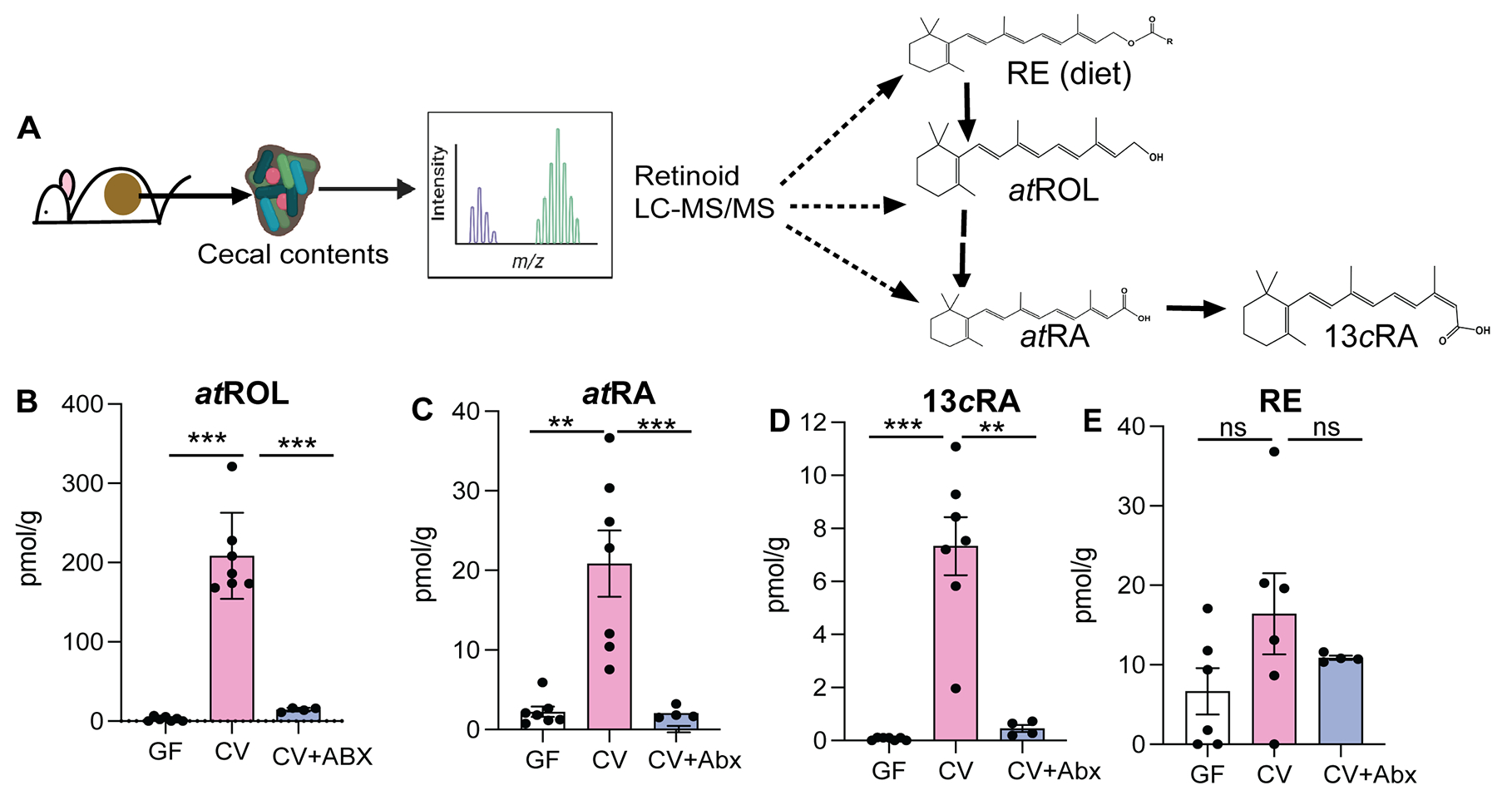
"Next, we investigated if VA metabolic activity observed in the gut lumen was correlated with presence of specific bacterial members. To determine the VA metabolic potential of different bacterial groups, CV mice were treated with either
- Vancomycin, which targets anaerobic commensals belonging to class Clostridia and Bacteroidia (Duncan et al., 2021), or
- Neomycin, which specifically targets aerobic commensals such as Proteobacteria (Hazenberg et al., 1983).
We observed that vancomycin treated mice had significantly lower ROL, atRA and 13cisRA concentrations in the gut lumen compared to untreated mice (
Figures 2A–C). Neomycin treatment did not affect retinoid levels in the gut lumen. The reduction in retinoids in the gut lumen of vancomycin treated mice was not due to reduced bacterial load (Figure S2C) suggesting that vancomycin sensitive commensal anaerobes provide the VA metabolic activity in the gut lumen.""To confirm the differential ability of commensal bacteria to metabolize VA, we colonized GF mice with Proteobacteria that bloom during vancomycin treatment (
Figure 2D). A second group of GF mice were given Clostridia spores in addition to Proteobacteria isolated from vancomycin treated mice. GF mice colonized with Proteobacteria alone did not show any increase and had comparable amounts of retinoids in the gut lumen as GF controls (Figures 2E–G). In contrast GF mice colonized with Proteobacteria and Clostridia spores showed significant increase in concentrations of atROL, atRA and 13cisRA in the gut lumen. 16S rRNA analysis of cecal community confirmed that gut microbiomes of Proteobacteria colonized mice lacked commensal anaerobes while mice that received spores in addition to Proteobacteria had high abundance of bacteria belonging to class Clostridia that restored all three retinoids in the gut lumen (Figure 2H).""We previously showed that gut bacteria can modulate VA metabolic machinery in the IECs and regulate RA amounts in intestinal tissue (Grizotte-Lake et al., 2018). To establish that bacteria intrinsic VA metabolic activity in the gut lumen is independent of host VA metabolic machinery, cecal contents from CV mice were harvested and grown in nutrients rich media and then supplemented with 10mM RE or ROL under anaerobic conditions. After 3 hrs., conversion of VA precursors into ROL, atRA and 13cisRA was quantified by LC-MS/MS (
Figure 3A). In the presence of bacteria, RE was hydrolyzed into ROL and ROL was oxidized to RA isomers (Figures 3B–F and S3). Bacterial cultures by themselves did not contain any detectable ROL or RE. The media alone supplemented with RE, or ROL did not yield quantifiable metabolites (Figures 3B–F) suggesting that there was no spontaneous conversion of RE and ROL into RA. These results establish that cecal bacteria can metabolize dietary VA precursors to active metabolites independent of the mammalian hosts and that gut bacteria encode the genetic machinery needed for performing VA metabolism." "Taken together, our results demonstrate that culturable consortia of gut bacteria can perform all the enzymatic reactions required for VA metabolism generating retinoid metabolites independently of the host.""Many Lactobacillus spp are naturally resistant to vancomycin (Swenson et al., 1990), and accordingly we observe that vancomycin treatment can result in bloom of Lactobacillus spp (
Figure S2B). We found that L. intestinalis, which has high ALDH activity was highly sensitive to vancomycin (Figure S4B) suggesting that loss of L. intestinalis upon vancomycin treatment could result in reduced retinoid metabolites in the gut. To evaluate if probiotic application of L. intestinalis to mice during vancomycin treatment would restore RA levels in the gut, mice were treated for 10 days with vancomycin and were given three applications of 108 CFUs of L. intestinalis or L. murinus. Similar bacterial loads were observed in all groups (Figure S4C). Retinoid analysis showed that L. intestinalis restored atRA levels of vancomycin treated mice while L. murinus, that does not show ALDH activity could not restore gut RA levels in vancomycin treated mice (Figure 4F)."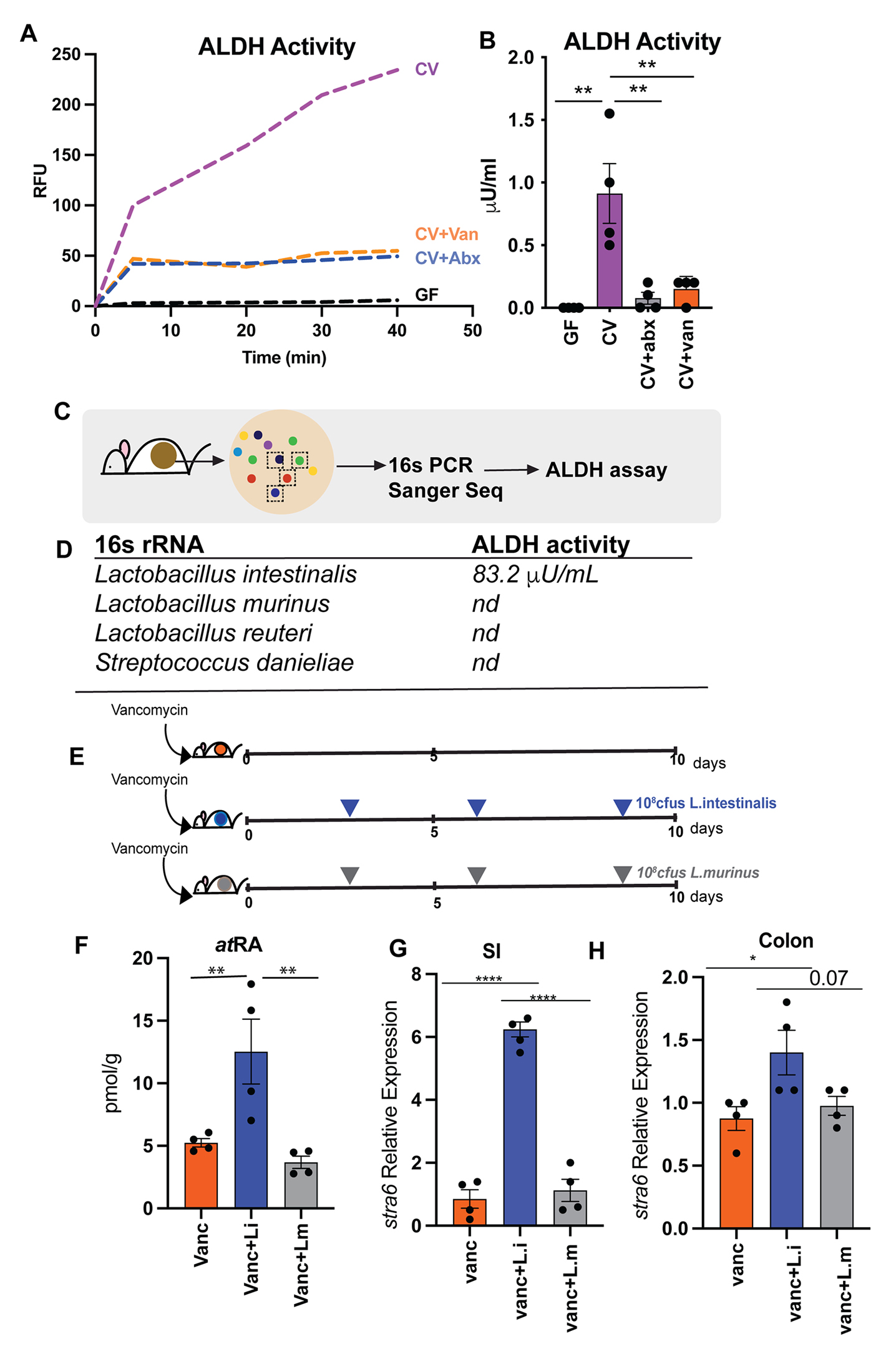
"[..]monocolonization of GF mice with either L. intestinalis, SFB or B. bifidum did not restore RA levels in the gut of GF mice (
Figure S4A) indicating that VA metabolism is an emergent property of gut microbiome and requires other members of consortia to participate in multistep conversion of VA to RA. In vancomycin treated animals L.intestinalis is able to restore RA production due to presence of vancomycin resistant bacteria that provide the metabolic machinery needed for other steps of VA metabolic pathway.""Next, we wanted to determine if restoration of RA in gut lumen following L. intestinalis treatment would induce RA responsive genes in the host. To do this we quantified known RA inducible host gene, Stra6 (Carrera et al., 2013; Kawaguchi et al., 2007). STRA6, has been shown to be essential in facilitating the cellular entry and exit of retinol and therefore is key in regulating how much retinol host cell can uptake. RA inducible gene stra6 was significantly induced in small intestine and colon of L. intestinalis supplemented mice compared to non-supplemented and mice gavaged with L. murinis (Figures 4G–H). We also observed higher expression of Nos2 gene [iNOS] in small intestine of the L. intestinalis supplemented mice in comparison to non-treated and L. murinis supplemented mice (
Figure S4D). The nitric oxide synthase 2 (nos2) gene was recently shown to be inducible by bacteria derived RA in the intestinal epithelium and its upregulation protected against pathogen colonization (Woo et al., 2021). We did not detect any significant differences in the pattern of inflammatory cytokines expressions in the small intestine and colon between the groups (Figures S4E–F) suggesting that addition of L. intestinalis is not linked to inflammation. Overall, our results demonstrate that bacteria with ALDH activity can restore RA following its depletion upon antibiotic treatment and promote the induction of RA responsive genes in the host." -
RE: Resources for authorsposted in Bioenergetic Development
Two months of sustained routine to form a habit is the median time:
How are habits formed: Modelling habit formation in the real world
"
Table 2shows the values for the curve parameters for the 39 participants for whom the model was a good fit, andFigure 3shows examples of the modelled curves of their data. Ten of these participants were performing eating behaviours, 15 drinking behaviours and 13 exercise behaviours. The median time to reach 95% of asymptote was 66 days, with a range from 18 to 254 days."
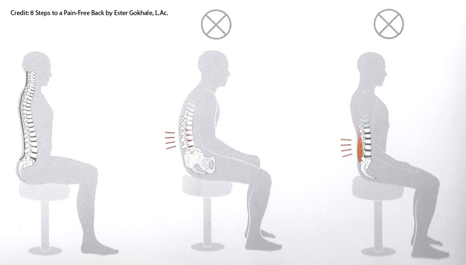
Racing chairs race you to the orthopedist:

For people who read a script while looking at the camera, an alternative would be to disappear from the scene to give space to supportive content on the screen, but appear at the beginning to greet and end to conclude if you want to keep it personal.
Narration may be viewed as a means to invite your victims next to you to show them your perspective rather than confronting them to defend your ideas.

-
RE: Resources for authorsposted in Bioenergetic Development
@jwayne
People usually lean forward to write: talk about being inclined versus disinclined. Assuming that you don't write on your ceiling, looking down must promote focus on the task, as it tends to isolate you from the surroundings. The head angle change might also have minor effects on circulation that can be significant over a long period, but I never stumbled upon any information on this. Perhaps spacecraft designers have considered this aspect because they have to decide what to prioritize on interfaces and the consequences of placing displays on relegated spots.
Might interest you:
- The Pen Is Mightier Than the Keyboard: Advantages of Longhand Over Laptop Note Taking
- Don’t Ditch the Laptop Just Yet: A Direct Replication of Mueller and Oppenheimer’s (2014) Study 1 Plus Mini Meta-Analyses Across Similar Studies
- Is typewriting more resources‑demanding than handwriting in undergraduate students?
- The effect of note-taking modality on offloading and memory under cognitive load
-
RE: Conditional problems with vitamin A: a place for sane discussionsposted in Literature Review

It directs to..
a) Professor Smith:
"If a person takes in more toxic "vit" A than they excrete on a regular basis, then they are ACCUMULATING it in their liver and bodyfat
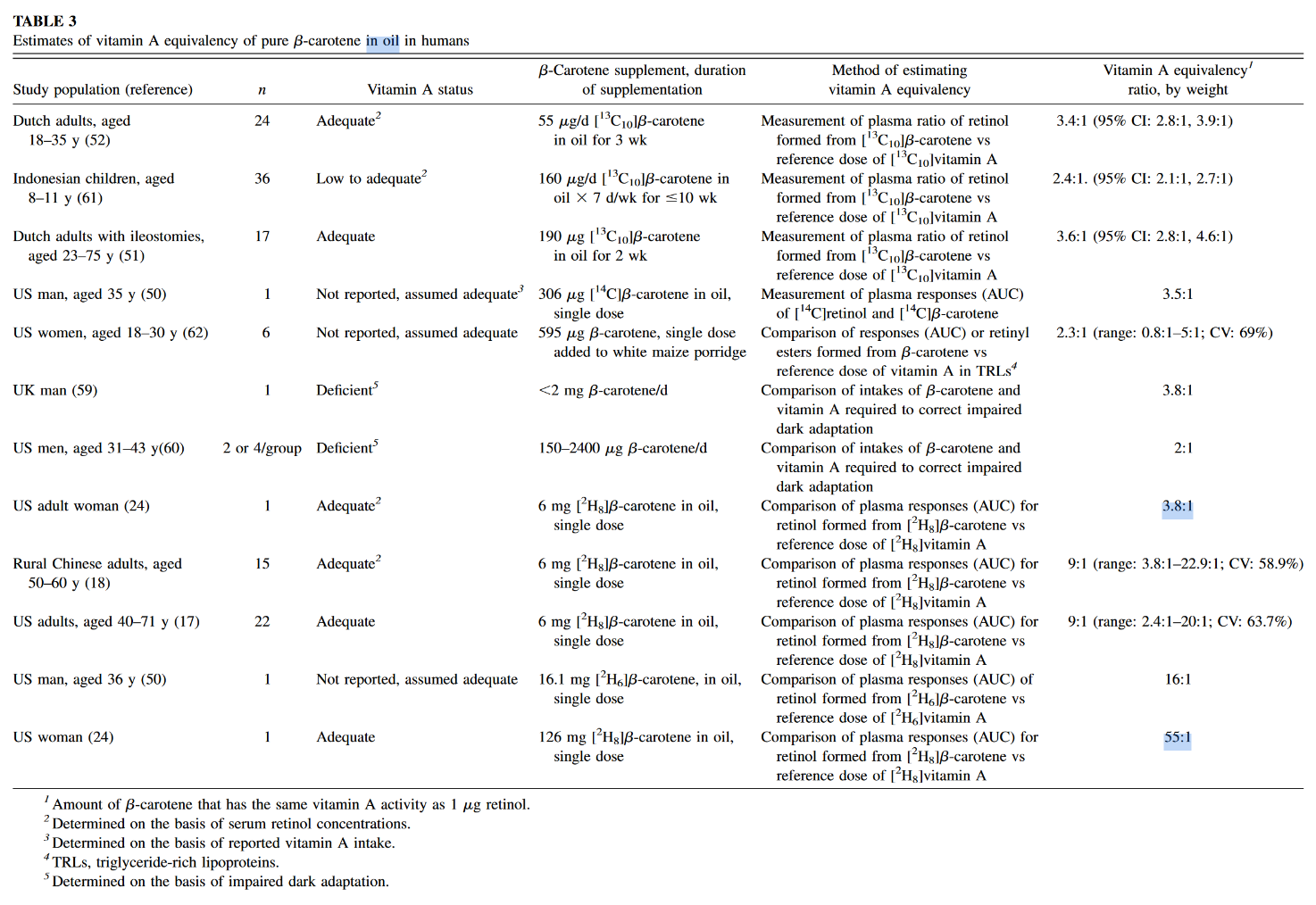
If 306 mcg beta-carotene (that's 1020 IU, one carrot has 6000 IU!) took 58 DAYS to turn over...do you realize just how fast you are filling up with this toxin?
1 beta-carotene eventually has to split into 2 retinaldehydes"
b) Disciple:
"Wow.
And this is also a great study for showing how much beta-carotene is actually converted to retinol. They write a minimum of 62% is turned into retinol. So a lot will eventually go the retinol route.
"What is clear is that, by our estimates, a minimum of 62% of the absorbed β-carotene was cleaved to vitamin A and by this reasoning the vitamin A value of β-carotene dose was 0.53. [The calculation is as follows: Assuming central cleavage, 1 mol of β-carotene yields 2 mol of vitamin A. If 62% of the absorbed β-carotene were cleaved to vitamin A, 1 mol of β-carotene would yield 1.24 mol of vitamin A. Thus, the vitamin A value of the absorbed dose is 1.24. Yet, the dose was 42.6% absorbed. So, the actual vitamin A value is (1.24) (0.426) or 0.53.]"
1 molecule of beta-carotene turns into 2 molecules of "active vitamin A".
The 0.53 value means that 1mg of ingested beta-carotene will turn into 0.5mg retinol. In other words, one medium carrot (eaten with lots of fat) can be equivalent to 27g beef liver.
Due to the time-delayed conversion, an acute poisoning is not possible, but a chronic poisoning is."
Their claims:
- a) "[..]one carrot has 6000 IU!" (1,800 mcg)
- b) "[..]one medium carrot (eaten with lots of fat) can be equivalent to 27g beef liver."
Beef liver:
- Poisonol 10,000 mcg/100 g
Carrot:
- Alpha-carotene: 2,000 mcg/carrot
- Beta-carotene: 5,000 mcg/carrot
We can disconsider half of the alpha-carotene amount and treat the other half as beta-carotene:
Beef liver:
- Poisonol 10,000 mcg/100 g
Carrot:
- Beta-carotene: 6,000 mcg/carrot
The author of the second post seems to be into the topic of poison A for a while, yet for some reason acts as if the information presented in the mentioned experiment was novel. These equivalences are found in almost all textbooks, even the main sections of the dedicated Wikipedia page have them, suggesting that pure b-macabrotene has half of the activity of poisonol:
- 1 µg RAE = 1 µg retinol from food or supplements
- 1 µg RAE = 2 µg all-trans-β-carotene from supplements
Now the equivalences from their claims:
- a) 1 µg RAE = 3.3 µg of all-trans-β-carotene from carrots
- b) 1 µg RAE = 2 µg of all-trans-β-carotene from carrots
The disciple is basing his equivalence on the following values from the linked experiment:
- Absorption: 42.6%
- Conversion: 62%
- Total (42.6% × 62%): 26%
This is close to a quarter, that results in a mass equivalence of about poison 1:4 b-mac, not 1:2 as he's arguing.
The source of confusion is because he's applying a molar to a mass ratio. B-carotene can yield two poisonols, but the mass is going to remain similar after cleavage and metabolism, with minor gains from the incorporation of new elements (that I'm going to disregard).
To arrive on his value:
- 26% × 2 = 53% (0.53)
It would've been preferable for the disciple to state:
"The 0.53 value means that 1 mg of ingested beta-carotene will turn into
0.5mg~0.25 mg retinol." The factor accounted for the potential of b-carotene to yield two poisonols, so it has to be applied after we halve the amount.At this stage, we can already discard the idea that a carrot will poison you as an ounce beef liver.
The spinach used in the quoted experiment served to tag macabrotene, the plant assimilated the labeled carbon of CO2 from the environment, allowing them to track the poison. But when it was time to contaminate the victim, the macabrotene was isolated and given in purified form at a low dose.
The food matrix lowers the availability of toxins, making them more difficult to be extracted, which is why researchers define a marked drop in efficiency when the b-macabrotene is derived from foods:
- 1 µg RAE = 1 µg retinol from food or supplements
- 1 µg RAE = 2 µg all-trans-β-carotene from supplements
- 1 µg RAE = 12 µg of all-trans-β-carotene from food
If we ignore this factor and pretend that fibrous carrots are an oil, the amount used in the experiment was modest (300 mcg) and we know that the rate of absorption and conversion tends to decrease as the dose gets high.
But let's reject this too and assume that it remains uninhibited up to 6 mg or so. However, the adjustment is beyond the meal content, the degree of body contamination with the poison is another factor that determines its fate. It's why the purity of the victim matters to interpret the dosing response to carotenoids: contaminated and decontaminated persons will metabolize it differently. When the body is overloaded, the rate of conversion to retinoids is reduced. To a lesser extent, the same goes for their absorption. Yet, carotenoids can be excreted intact.
"1 beta-carotene eventually has to split into 2 retinaldehydes"
It doesn't. His own equivalence conflicts with this claim, even if we discount the unabsorbed fraction (and he probably didn't):
"1 beta-carotene eventually has to split into 2 retinaldehydes":
- 6,000 mcg b-carotene → about 6,000 mcg PAE
"1 [absorbed] beta-carotene eventually has to split into 2 retinaldehydes":
- 6,000 mcg b-carotene → about 2,500 mcg PAE
"[..]one carrot has 6000 IU!":
- 6,000 mcg b-carotene → about 1,800 mcg PAE
Which is it?
"If you take something more than you can excrete, you accumulate"
Of course.
"If 306 mcg beta-carotene (that's 1020 IU, one carrot has 6000 IU!) took 58 DAYS to turn over...do you realize just how fast you are filling up with this toxin?"
A long half-life doesn't result in an indefinite accumulation, only until a steady state is reached, when poisoning for profits should plateau with regular consumption. Something like this in dosing more often:
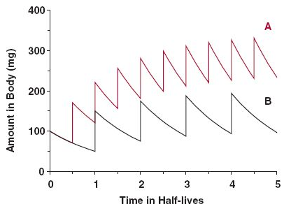
Source: the internet.Some accumulation is normal and healthy; or should we treat macabrotenoids as toxins to be nuked from the body at all costs? "Vitamin" E is a toxin too (Smith, 2023), but is it harmful when it's not detoxified fast enough to avoid a detectable level, leading to 'accumulation' to be associated with problems?
Consider this: you consume a meal, whose elimination would take 24 hours. Instead of waiting for this period to eat the next, you consume multiple meals in between (may be 4 in a day), that result in accumulation of matter, but without issues. The accumulation is temporary, purposeful and the body is adapted to it. It's not immediate, but the matter eventually leaves the body to the extent that it's consumed: no stuffing until explosion occurs because the input matches the output despite the delay.
Excess carotenoids or calories should come with signs. The early stages are benign, will indicate that the person has to cut back or improve the capacity to handle them. Yet, carotenemia isn't reliable to diagnose poison A toxicity because it can be caused for different reasons (such as lack of nutrients or poor protein synthesis). As an example, if a person needs to take supplemental thyroid hormones and suddenly stops the treatment, it can be followed by carotenemia.
The rate of storage and mobilization is adaptable as well. In case someone discontinues the consumption of poisons altogether, it can signal conservation and a longer persistence in the body. Therefore, the length is variable.
All in all, while we have people debating whether an inability to convert macabrotenes is common or a real concern for persons who don't consume preformed poisons, we have the poison A crowd alarming you that regular carrot consumption is a ticket to the hospital over time, for having the same potential of destruction as liver in the diet, neglecting the multiple steps that regulate these toxins as drugs. #toxicbileapocalypse
-
Mike Fave is on fireposted in Bioenergetics Discussion

Many of you enjoyed the posts of Miguel Favo, member CLASH of the Ray Peat Forum. He has reformulated his approach to increase his reach.
He now has two channels on YouTube, one for distilled content and the other for dissection with a touch of religion. Both are good, check them out.
If I'm not wrong, he has a newsletter and an ebook as well.
Miguel will soon take off. If you want to communicate with him before he turns into a posh celebrity, you're short of time.
He respects the audience and doesn't resort to cheap tactics to promote his content. If you like his material, share with others to speed up his popularization.
Maybe harass Mexico and Bulgaria to request their collaboration in The Generative Energy podcast too.
-
Details about magnesium absorptionposted in Literature Review
Challenges in the Diagnosis of Magnesium Status
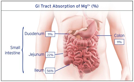
"Due to the complex nature of magnesium absorption, segments of the GI tract can vary in their contribution to absorption; however, under normal physiologic conditions the general guide is the duodenum absorbs 11%, the jejunum 22%, the ileum 56%, and the colon 11% (Figure 3) [3,58]."
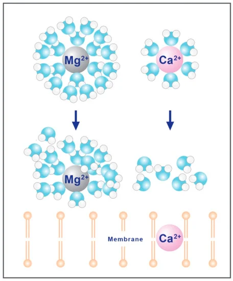
ㅤ"Magnesium is a divalent cation, which plays a critical role in how the mineral is absorbed [83,84]. Magnesium is the most densely charged of all the biological cations, due to a high charge to radius ratio, resulting in high hydration energy for the Mg2+ cation [83]. This hydration energy results in tight coordination with a double layer of water molecules, increasing the hydrodynamic radius 400 times that of the dehydrated radius [83,85], resulting in an aquated cation that is too large to transverse typical ion channels (Figure 5) [86]. The removal of the hydration shell around magnesium is a precondition for absorption and can be accomplished by both TRPM6 and TRPM7 [⇈] and the magnesium associated paracellular claudins [64,67,87,88]."
It can be presumed that low doses would skew the absorption site distribution towards the upper parts, but it's counterintuitive: the active process of magnesium absorption, that prevails at lower intakes, occurs primarily farther down the small intestine.
Segmental transport of Ca2+ and Mg2+ along the gastrointestinal tract
"Although occasionally debated, paracellular absorption is generally considered to be predominant under normal physiological circumstances (3, 4, 32, 40, 44). When paracellular absorption cannot meet the daily need of the body, transcellular absorption becomes essential (6, 40). Specific transport proteins, located in the luminal and basolateral membrane of the epithelial cells, govern this latter process (23, 26, 32)."
"Transcellular absorption of Ca2+ and Mg2+ from the intestinal lumen is facilitated by members of the transient receptor potential (TRP) ion channel family, which represents the rate-limiting factor in the transcellular transport process. Intestinal
absorption of Ca2+ is mediated via TRP vanilloid type 6 (TRPV6), whereasabsorption of Mg2+ takes place via TRP melastatin type 6 (TRPM6) (20, 30, 43)." "For transcellular Mg2+ transport in the intestine TRPM7 and cyclin M4 (CNNM4) seem to be important in addition to TRPM6, since they are though to play a role in apical and basolateral transport of Mg2+, respectively (38, 47).""TRPM6 is expressed in the distal regions of the gastrointestinal tract of human subjects and mice (Fig. 2B). Interestingly, like TRPV6, TRPM6 in mice is expressed strongly in the cecum (Fig. 2B)."
"TRPM7 was evenly expressed throughout the gastrointestinal tract (Fig. 2D). Expression of the basolateral Mg2+ transporter CNNM4 was most prominent in the distal gastrointestinal segments, resembling the expression profile of TRPM6 (Fig. 2F). However, in contrast to TRPM6, CNNM4 expression was not completely absent in the proximal gastrointestinal tract."
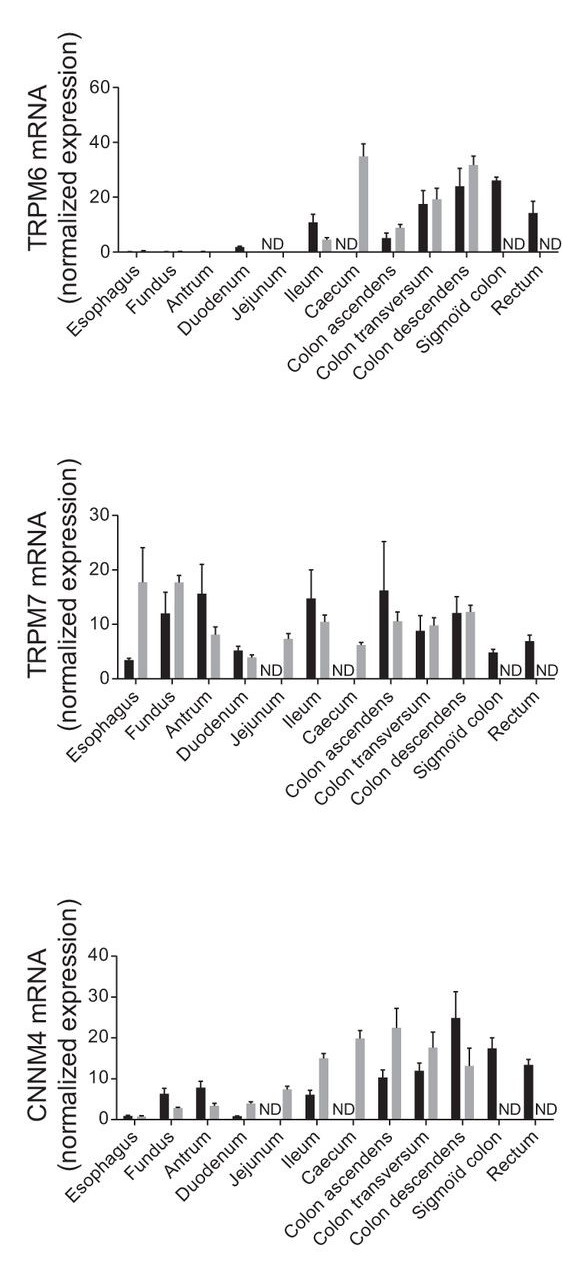
- Dark bars: human
- Gray bars: mice
ㅤ
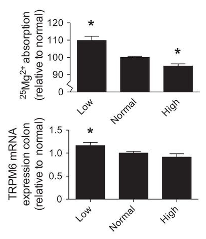
"To investigate whether Mg2+ absorption is regulated by dietary Mg2+ content, mice were fed a diet with a low (0.02% wt/wt Mg2+), a normal (0.25% wt/wt Mg2+), or a high (0.48% wt/wt Mg2+) Mg2+ content for 2 wk."
"Urinary excretion of Na+, K+, and Cl− did not differ significantly between the groups, excretion of Pi was significantly higher in the low-Mg2+-diet group compared with the groups receiving normal and high-Mg2+ diets (
Table 2).""Dietary Mg2+ restriction significantly stimulated the rate of intestinal absorption of Mg2+ (Fig. 3G). Conversely, intestinal absorption of Mg2+ in the high-Mg2+-diet group was diminished compared with the normal group. The absorption of Mg2+ in the low-Mg2+-diet group was significantly higher compared with the normal group (110.0 ± 3.0% and 100.0 ± 0.1%, respectively, P < 0.01) and in the group on the high-Mg2+ diet absorption was significantly lower compared with the normal group (95.0 ± 1.0 vs. 100.0 ± 0.1%, respectively, P < 0.05) (Fig. 3H). TRPM6 mRNA expression levels in the colon were slightly, but significantly, regulated by dietary Mg2+, showing an increase in the group on the low-Mg2+ diet and a decrease as a consequence of the high-Mg2+ diet (Fig. 3I)."
ㅤ"The proximal Mg2+ absorption was higher compared with the distal part of the intestine at a high luminal concentration of Mg2+ (44 mM) (Fig. 4E), but not at lower luminal concentrations (1.6 and 10 mM Mg2+) (Fig. 4, A and C). The relationship between the absorption of 25Mg2+ and the luminal Mg2+ concentration is displayed in Fig. 4G. In the proximal intestine, the absorption of 25Mg2+ resembles a linear relation to the buffer Mg2+ concentration, whereas in the distal part of the intestine the absorption of Mg2+ seems saturable at higher Mg2+ concentrations."
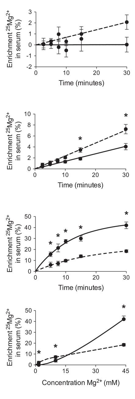
"Paracellular and transcellular absorption of Mg2+ in intestinal segments. Solid lines represent mice with a cannula placed in the stomach, dashed lines represent mice with a cannula placed in the cecum. 25Mg2+ absorption by using a buffer containing 1.6 (A), 10 (C), or 44 (E) mM Mg2+." "Relationship between luminal Mg2+ concentration and 25Mg2+ absorption (G)."
"The findings in our study, therefore, suggest that
transcellular Ca2+ absorption takes place in the proximal region of the intestine, whereastranscellular Mg2+ absorption mainly occurs in the distal gastrointestinal segments." "Paracellular absorption of Ca2+ and Mg2+ mainly takes place in the duodenum and jejunum."
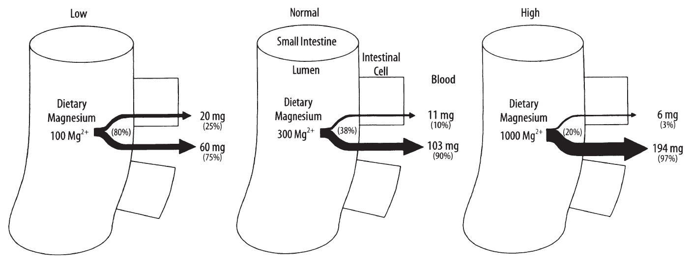
A representation of how much of the daily amount is absorbed and the route distribution (transcellular or through the cell; paracellular or between cells):
Intestinal absorption of magnesium
-
Vitamin C in the prevention of cellular respiration (CcO) inhibition by sulfideposted in Literature Review
Modulation of sulfide oxidation and toxicity in rat mitochondria by dehydroascorbic acid
"As isolated cytochrome c oxidase activity is strongly inhibited by sulfide at concentrations lower than 1 μM, this aspect has to be considered physiologically relevant even under healthy conditions [20]. Interestingly, respiration of isolated mitochondria or whole cells is significantly less susceptible and not affected by sulfide concentrations up to 5–20 μM [19,21, this study]. At least partially this discrepancy can be explained by an efficient detoxification of the sulfide by the mitochondrial oxidation pathway, which rapidly lowers the ambient concentration. Moreover, other cellular factors might be present in vivo to control the binding of sulfide to cytochrome c oxidase."
"Dehydroascorbic acid is a good candidate for such modifying factor because it has been shown to affect sulfide oxidation in the sulfide adapted invertebrate Arenicola marina [9], and it is readily available in mammalian mitochondria. Ascorbic acid, the main form of vitamin c in cells, functions as a reductant during the biosynthesis of collagen, carnitine and catecholamine thereby producing DHA [22]. Rat and mouse liver contains about 3 mM ascorbic acid [23,24], but it is difficult to determine the physiological DHA concentration because it is rapidly recycled to ascorbic acid by enzymatic and non-enzymatic reduction. A fraction of 5–7 % DHA has been estimated for plasma [25]. However, DHA concentrations can be markedly increased under certain conditions involving oxidative stress such as inflammation or ischemia reperfusion injury [22]. Especially in mitochondria local gradients might occur with a transient increase of DHA in regions of high ROS production at the respiratory chain, where ascorbic acid acts as a major antioxidant. Furthermore, ascorbic acid transport into mitochondria mainly takes place in form of DHA via the glucose transporter GLUT1 [26]."
"The results presented here demonstrate that DHA is able to completely prevent inhibition of cytochrome c oxidase at low sulfide concentrations, which are certainly relevant for mitochondria in the physiological context. As a consequence, mitochondria maintain their regular energy metabolism including ATP production in the presence of this potent inhibitor of the respiratory chain. Partial protection is provided even at relatively high sulfide concentrations that might occur locally or temporarily."
"The easiest explanation for the decrease of sulfide toxicity in the presence of DHA would be a direct interaction of the two compounds. Very high concentrations of sulfide, i.e. a solution saturated with H2S gas, can be used as a reductant to quantitatively convert DHA to ascorbate [27]. Rates of non-enzymatic sulfide consumption also increased after the addition of DHA under the experimental conditions used in this study. Therefore, a decrease of the free sulfide concentration by binding to DHA and chemical oxidation is certainly part of the story. But it is not sufficient to explain all results. If the effect was exclusively chemical it could be expected to be independent of the assay system used to determine cytochrome c oxidase activity. Nevertheless respiration with different substrates such as succinate, NADH and TMPD continued in the presence of 50 μM sulfide plus 1 mM DHA whereas the oxidation of externally added cytochrome c was already completely inhibited at lower sulfide concentrations. Therefore, an additional mechanism probably exists that prevents binding of sulfide to cytochrome c oxidase either by decreasing the affinity or via steric protection of the active site of the enzyme. This effect is independent of membrane potential, as it was not abolished by freezing and thawing of the mitochondria (data not shown)."
"Obviously the rapid oxidation to non-toxic compounds is important for an efficient detoxification of exogenous as well as endogenous sulfide. Especially in the liver this pathway is involved in the degradation of the sulfur containing amino acids methionine and cysteine [42]. DHA also decreases sulfide toxicity in mitochondria from other tissues that have been shown to oxidize sulfide enzymatically such as kidney and heart [19]. As a consequence, sulfide can be used as an inorganic substrate for ATP production in a wide concentration range, but this aspect is presumably of limited importance compared to the normal organic substrates. In contrast to most tissues, brain mitochondria probably lack at least part of the sulfide oxidizing pathway and are therefore more sensitive to sulfide poisoning [19]. The present data support this finding as even in the presence of DHA no enzymatic sulfide oxidation was detectable. However, the protective effect of DHA was most pronounced in the brain demonstrating that this mechanism is particularly important in the absence of an efficient sulfide detoxification pathway."
"The identification of DHA as an instrument to reduce sulfide toxicity may become clinically relevant during the treatment of sulfide poisoning either after accidental environmental exposure or in patients that accumulate endogenous sulfide, e.g. in ethylmalonic encephalopathy. This directed amplification of sulfide oxidation rates could be an alternative therapeutic approach to the inhibition of sulfide producing enzymes, which is already being tested [44]. Maximal intracellular concentrations of vitamin C (3–4 mM) are achieved by supplementation of 200 mg ascorbic acid per day [45]. For acute treatment it is also possible to administer DHA directly via intravenous injection, which raises intracellular ascorbate concentrations to supraphysiological levels and thus probably provides enough DHA to be protective against sulfide toxicity [46]."
-
RE: A list of members banned from the Ray Peat Forumposted in The Junkyard
@saturnuscv said in A list of members banned from the Ray Peat Forum:
User “feedandseed” was banned in the original haidut ban thread
Melissa would've jumped in to intervene if she wasn't busy legitimizing Jorge's attacker. Her superpowers are brought up when convenient, but soon defaults to filing her nails while accepting the sportive disposal of members.
@TheSir said in A list of members banned from the Ray Peat Forum:
I am now banned too as a result of this post.
I've stumbled upon some of your questionings, they were precise and exemplary. It's a loss for them.
@deliciousfruit said in A list of members banned from the Ray Peat Forum:
Got banned for saying this:

I wasn't even trolling, I've been taking 15mg of copper daily for months and have only seen benefits.I guess having an experience antithetical to the forum owner's narrative is a bannable offense now.
It's a little risky, but shows the concern that they have for members: you're taking something that they regard as harmful and you're dumped to continue to do it elsewhere.
@Sweet-Meat said in A list of members banned from the Ray Peat Forum:
did he ban anyone paying subscriptions?
Yes. What hasn't been banned yet?

-
Pantothenic acid forms in foodsposted in Literature Review
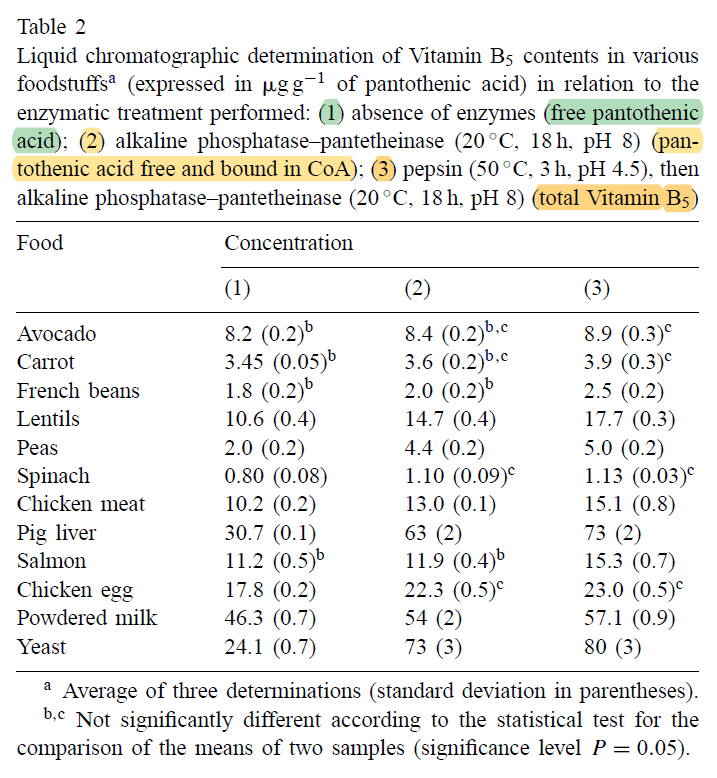
"[..]CoA is the major pantothenic acid-containing compound in yeast, pig liver and peas, but that it was not found, on the other hand, in the avocado, carrots, French beans and salmon. In the four other non-supplemented foodstuffs studied (chicken egg, chicken meat, spinach, lentils), the percentage of pantothenic acid released from CoA is included between 20 and 30%. These results confirm that CoA is effectively the major pantothenic acid-containing compound in animal liver and yeast as has already been mentioned by Brown [29] and Gonthier et al. [6], but they seem partially to refute the general but unsupported allegation of Ball [30], according to which the majority of naturally occurring pantothenic acid would be in the form of CoA."
Liver Coenzyme A Levels in the Vitamin B6-Deficient Rat
"Liver CoA concentration in the rat was decreased 50% by vitamin B6 deficiency and 66% by pantothenic acid deficiency. The decrease caused by vitamin B6 deficiency was independent of food intake and was not remedied by cystine supplementation of a 20% casein diet."
-
RE: The resynthesis of methionine through betaine (BHMT) is confined to the liver and kidneysposted in Literature Review
"Betaine-homocysteine S-methyltransferase (BHMT) is the only enzyme known to catabolize betaine. In addition to being a substrate for BHMT, betaine also functions as an osmoprotectant that accumulates in the kidney medulla under conditions of high extracellular osmolarity. The mechanisms that regulate the partitioning of betaine between its use as a methyl donor and its accumulation as an osmoprotectant are not completely understood. The aim of this study was to determine whether BHMT expression is regulated by salt intake. This report shows that guinea pigs express BHMT in the liver, kidney, and pancreas and that the steady-state levels of BHMT mRNA in kidney and liver decrease 68% and 93% in guinea pigs consuming tap water containing high levels of salt compared with animals provided untreated tap water. The animals consuming the salt water also had ∼50% less BHMT activity in the liver and kidney, and steady-state protein levels decreased ∼30% in both organs. Pancreatic BHMT activity and protein levels were unaffected by the high salt treatment. The complex mechanisms involved in the downregulation of hepatic and renal BHMT expression in guinea pigs drinking salt water remain to be clarified, but the physiological significance of this downregulation may be to expedite the transport and accumulation of betaine into the kidney medulla under conditions of high extracellular osmolarity."
-
Nutrition in Small Intestinal Bacterial Overgrowthposted in Literature Review
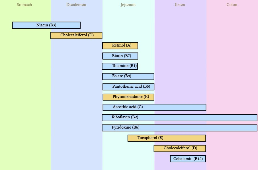
Intestinal absorption of micronutrients and macronutrients | Deranged Physiology
Small Intestinal Bacterial Overgrowth
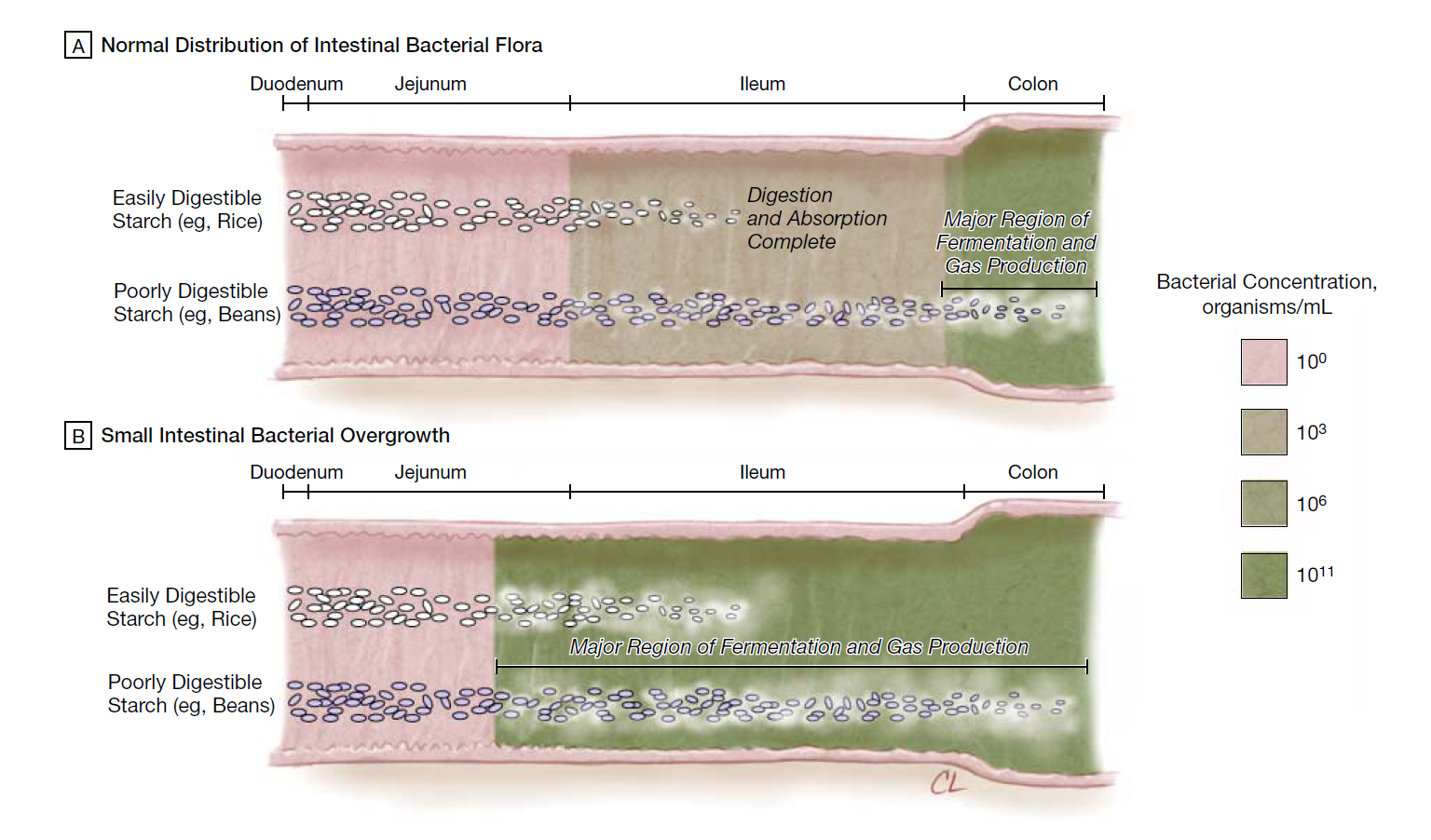
"In rats, experimentally induced SIBO leads to the appearance of gut bacteria in the mesenteric lymph nodes and visceral organs.[49] A potentially important consequence of bacterial translocation is immune activation. In a report of 11 patients, an increase in the number of intraepithelial lymphocytes was observed as mucosal evidence of this immune response to confirmed bacterial translocation.[53] This adverse outcome could explain why the normal gut has defensive mechanisms in place to keep the bacterial flora away from the small intestine, particularly the bowel proximal to the ileum."
"Nucera et al[37] found a high rate of disappearance of malabsorption to lactose (86.6%), fructose (97.5%), and sorbitol (90.9%) once SIBO was eradicated."
-
Zinc supplementationposted in Literature Review
"[..]the chemical form of zinc (salt or chelate), the pharmaceutical formulation (presence of excipients), and the division of the administered dose are other factors that modify the oral bioavailability of zinc [18]. Considering the pharmaceutical formulation, Neve et al [18] noted a decrease in the oral bioavailability of zinc when zinc gluconate was associated with starch, lactose, hydrated silica, and magnesium stearate (excipients present in Rubozinc), compared with zinc gluconate without excipients."
"In the case of our results, the significantly higher oral bioavailability of zinc when administered as bis-glycinate compared with gluconate is not surprising, given that bis- glycinate enhances the oral bioavailability of others ions, particularly iron."
"The higher absorption of zinc when chelated with bis-glycinate may well be explained by its chemical structure [27].
- Firstly, each glycinate molecule contains two functional groups that are capable of entering into covalent and coordinate covalent bonds with zinc.
- Secondly, each one can create a ring structure with the zinc ion and form a sterically and energetically stable structure.
- Thirdly, zinc bis-glycinate has a low molecular weight (< 1000 daltons) and is able to cross cell membranes.
- Lastly, the bis-glycinate chelate is not affected by pH changes from 2 to 6 [28].
Consequently, bis-glycinate may protect a significant fraction of the zinc from interactions with lumenal gastro-intestinal components, dietary components, and drugs [29] whereas in the current salts, the zinc may not be protected from such interactions."
Zinc Absorption Adapts to Zinc Supplementation in Postmenopausal Women
"To help establish common baseline conditions for all subjects, this double-blind study began with a 12-d pre-treatment period during which all subjects consumed the basal diet plus 6.0 mg Zn (as Zn gluconate) and 1.0 mg Cu (as Cu sulfate) daily. For the remaining 156 days, subjects continued the basal diets but with supplemental Cu reduced to 0.5 mg/d, and with random assignment of 9, 27 or 42 mg/d supplemental Zn, for total average Zn intakes of 14 (n = 6), 32 (n = 3), or 47 mg/d (n = 3). Both Zn and Cu supplements were divided into three equal portions, mixed with juice, and given with each of the three main meals, daily."
"Initially (wk 0) the women consuming 14, 32, and 47 mg Zn/d absorbed 4.6, 8.7, and 10.3 mg Zn (pooled SD = 1.3), respectively, suggesting a partial saturation of Zn absorption kinetics when Zn intakes met or exceeded 32 mg/d. However, with time, Zn absorption by those ingesting 32 and 47 mg Zn/d declined so that differences in total Zn absorbed were not significant at 8 wk (5.4, 5.8, 6.4 mg/d, respectively) and were negligible by 16 wk (5.0, 5.0, 5.1 mg/d)."
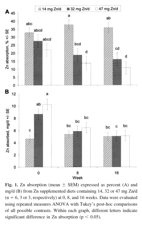
"The final absolute Zn absorption at 16 wk was [..] strikingly constant for all subjects, consistent with the overall finding that complete adaptation occurred (Fig. 1). Given the unusually extended period of highly controlled feeding conditions, and the precision of the whole body counting method, these significant results provide valuable information on human Zn homeostasis despite the small number of subjects."
"The complete absorptive adaptation observed in this study may be limited to adults with adequate Zn status who consume relatively low-phytate diets."