@greekdemigod looks like sperm cells are especially vulnerable , successful implantation apparently went down by half from just 1x a week for 30 minutes at 39 degrees, and might negatively affect offspring https://www.rbmojournal.com/article/S1472-6483(20)30073-0/abstract
20 minutes at 40 degrees water immersion too https://www.sciencedirect.com/science/article/abs/pii/S0093691X20305343
and in humans it would take 2-3 months at least to restore to normal after stopping because of regen time ,
human study, using extreme heat sauna 2x a week, impaired sperm, but didn't find negative effect on hormone production, motility back to normal by a few months after stopping but full count at 6 months
https://emilkirkegaard.dk/en/wp-content/uploads/Seminal-and-molecular-evidence-that-sauna-exposure-affects-human-spermatogenesis-.pdf
@Jakeandpace nice add with the icing idea, or just a bag of cold water to avoid other extreme (or if its hot enough i guess it doesnt matter)
I found it takes about 10 minutes to elevate body temp well and lasts a bit after stopping
@Mauritio
In type 2 diabetes, insulin resistance and increased glucose levels cause mitochondrial dysfunction and decreased expression of HSP60 in the brain, which promotes brain hyperglycemia, resulting in high ROS levels and contributing to mitochondrial dysfunction [94]. This highlights the importance of HSP60 expression in type 2 diabetes for maintaining mitochondrial proteostasis.
Notably, the interaction between HSP60 and toll-like receptors (TLRs) supports the hypothesis that HSP60 could be involved in disease progression. However, the dysregulation of HSP60 in mouse hypothalamic cell lines and brain samples from T2DM patients further supports its role in the pathophysiology of T2DM [95,96].
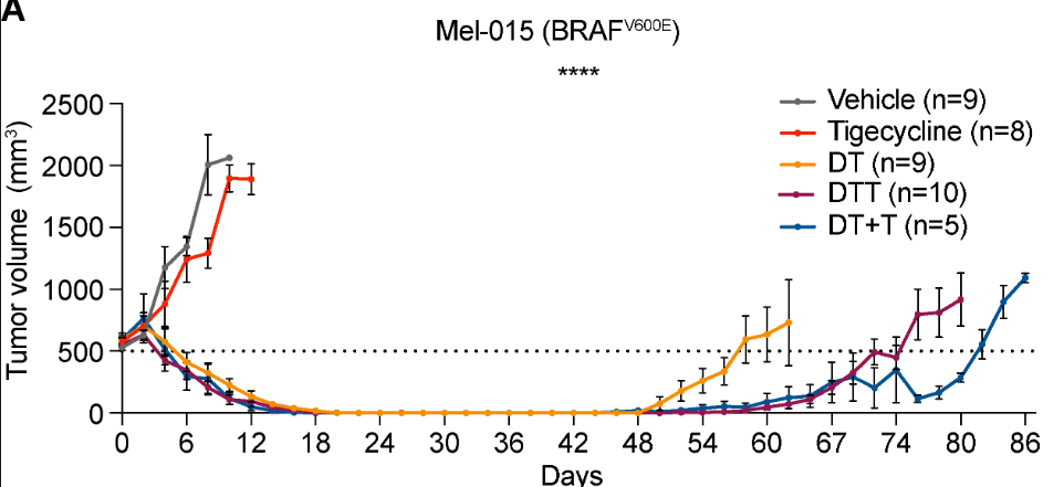
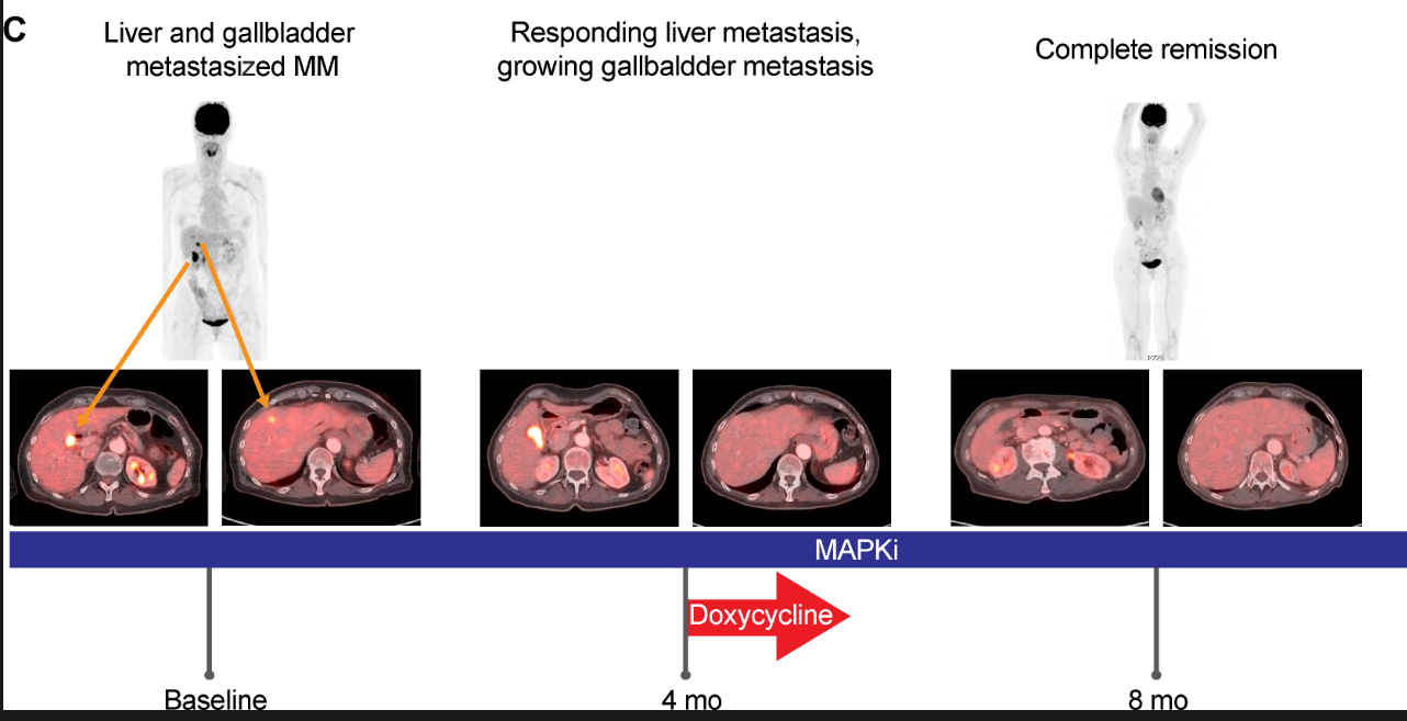
Yes and the pro-metabolic things lowering them: progesterone, tetracyclines, selenium, methylene blue, ...
aye rays take seems fair enough based on these, but some benefits regardless of elevation
HSP60 (majority found in mitochondria)
, Warning for using hsp60 inhibitors in cancer, over-expression of hsp60 (outside of heat induced) can promote cancer growth, but its also vital for healthy cells, removing HSP60 = mitochondria dysfunction + heart failure https://www.nature.com/articles/s41418-019-0374-x
Recently, it has been reported that in burn sepsis situations, mitochondrial energy metabolism is restored in hepatocytes through upregulation and activation of SIRT4, an NAD+-dependent deacetylase, which subsequently reduces acetylation of HSP60 and facilitates the assembly of the HSP60–HSP10 complex. This reduced acetylation of HSP60–HSP10 restores the mitochondrial energy metabolism at complex II and complex III via glutamine-mediated SIRT4 activity (Table 2). Therefore, HSP60 may serve as a target of SIRT4 in hepatocytes, improving ATP production and protecting from burn-sepsis-dependent organ injury
HSP70 inhibits steroidogenesis? Where HSP60 is pro steroidogenesis? (or at least pro progesterone production)
https://pubmed.ncbi.nlm.nih.gov/7582246/
We have described two mitochondrial (mt) myopathy patients with reduced activities of various mt enzymes associated with significantly decreased amounts of heat shock protein 60 (hsp60). Experimental evidence suggested that the lack of hsp60 was the primary defect. Since hsp60 is essential for the proper folding of enzyme subunits in the mt matrix a partial deficiency of this protein can explain the observed defects of the mitochondria.
Here we report on morphological studies aimed at obtaining more insight into the relation between lack of hsp60 and pathological changes of the mitochondria. Under standard culture conditions mitochondria in the partially hsp60 deficient fibroblasts showed profound morphological aberrations. In contrast, the mitochondria in fibroblasts from a MELAS patient and a cytochrome c oxidase-deficient patient appeared normal.
Under stress conditions the integrity of the hsp60 deficient mitochondria declined even further: heat shock induced a temporary collapse of the electrochemical potential across the inner mt membrane, but did not affect the ultrastructure of the mitochondria; prolonged growth in confluent cultures resulted in decrease in mt number.
The altered mt morphology in the hsp60 deficient cells is probably indicative of the severely impaired mt metabolism whereas the decreased stress tolerance is likely to be a direct result of paucity of the heat shock protein. Both variables are potentially useful in the diagnosis and molecular characterization of mt disorders with systemic manifestation and multiple enzyme deficiency.
https://pubmed.ncbi.nlm.nih.gov/7986829/
In a recent paper (Agsteribbe et al. (1993) Biochem. Biophys. Res. Commun. 193, 146-154) we suggested deficiency of heat shock protein 60 (hsp60) as the possible cause of a systemic mitochondrial encephalomyopathy with multiple deficiency of mitochondrial enzymes. In this paper we present new data which strongly support this hypothesis. Hsp60 deficiency appeared to be not a common side effect of impaired mitochondrial metabolism as eight out of ten fibroblast cultures from patients with systemic mitochondrial myopathy contained normal quantities of the protein.
https://www.academia.edu/72683004/Role_of_HSP60_in_Steroidogenesis_and_Reproduction
incorporation of specifc proteins at the level of mitochondria becomes relevant because most proteins that constitute them are encoded in the nucleus and are synthetized in the cytoplasm with a pre-sequence or a signal peptide that allows their transportation into mitochondria. This process requires complex systems, like the TIM and theTOM, where HSP are required (Marom et al. 2011). For example, for the incorporation of the translocator of adenine nucleotides into the inner mitochondrial membrane, the presence of Hsp60 is indispensable (Mahlke et al. 1990)
overexpression of Hsp70 inhibits the synthesis of steroid hormones in luteal cells. It has been suggested that the action mechanism by which Hsp70 inhibits steroids’ synthesis is by interfering with cholesterol translocation to the mitochondrial cyto-
chrome P450scc (Khanna et al. 1994). Results revealed that the presence of Hsp70 in Leydig cells was constant, whereas Hsp60 was detected in both germinal and Leydig cells associated with an increase in the heat shock factor
1 and a diminution in the StAR protein, with the consequent diminution in testosterone production (Oka et al. 2017)
Authors proposed that the heat shock factor 1 regulates testicular steroidogenesis by stabilizing cholesterol transport through StAR. the heat shock increases eight to ten times the synthesis of progesterone with respect to control cells. Authors suggest that one of the effects could be the modifcation of cholesterol transport at the mitochondrial level (Khanna et al. 1994, 1995a, 1995b).
It has also been reported that heat of the summer produces stress in bovines increasing infertility. In this sense it has been reported that all HSP increase, particularly Hsp60 increases 2.6 times, and Hsp70 type 3 was the highest with a 4.9-times increase. Although levels of StAR and P450scc diminished, this did not affect the synthesis of progesterone,
overexpression of Hsp60 in Leydig cells induced by human chorionic
gonadotropin and the follicle stimulating hormone is associated with the increase
in testosterone secretion and regulation of steroidogenesis (Miller 2013; Issop et al. 2013).
using MA-10 cells subjected to heat stress, where Hsp70 levels increased between 6 and 20 times, depending on the treatment time, the inhibition of steroidogenesis was induced, without affecting the P450scc or 3β-HSD activities;
It was proposed that RoA deletion produced important changes in the cell’s cytoskeleton, causing accumulation of lipid droplets, and avoiding mitochondrial fusion/fssion processes, thereby impeding steroidogenesis by decreasing mitochondrial Hsp60.
In follicular cells of the ovary, the induction of protein StAR synthesis was accom-
panied by an increase of Hsp60. Transfection with the whole electron transport chain of P450scc, which is needed for cholesterol transformation into pregnenolone, increased signifcantly the expression of Hsp60 with the concomitant hormonal increase (Monreal-Flores et al. 2017)
the interpretation was that the mitochondrial Hsp60 of the human placenta is directly related with the transport of cholesterol. Furthermore, the presence of
maleimides did not modify the oxidative phosphorylation in these mitochondria,
confrming that inhibition of progesterone synthesis is related with blocking the
active cysteines of Hsp60
a reproducible model of non-steroidogenic cells
(HEK293) transformed into steroidogenic with the transfection of P450scc system,
and the protein 3β-HSD confrmed that overexpression of Hsp60 increased the transformation of cholesterol into progesterone (Monreal-Flores et al. 2017). Consistent with these data, in vitro assays demonstrated that the recombinant His10-Hsp60 protein favored progesterone synthesis in mitochondria isolated from JEG-3 cells, confrming its participation in steroidogenesis
Hsp60 could bind cholesterol similarly to what has been reported for
the StAR protein in MA10 cells
In general, it has been reported that HSP are the frst proteins to be produced in in vitro culture of embryos, and that they are immunodominant antigens of some pathogens like Chlamydia trachomatis.
Similar results were obtained in women with Chlamydia trachomatis infections, in which high levels of anti-Hsp60 were related with a diminution in in vitro fertilization.
It has been proposed that the presence of HSP, in particular of Hsp70, could avoid the cytotoxic effects produced by TNFα, which induces activation of phospholipase A2 with the subsequent activation of mediators that induce inflammation and mitochondrial formation of reactive oxygen species
It is interesting that in the human placenta, Hsp60 is associated with the
steroidogenic process by participating in the transport of cholesterol at the mito-
chondrial level where is transformed into progesterone by the P450scc chain